Orbital tubercle (Latin: eminentia orbitalis)
Also called: Whitnall's tubercle; lateral orbital tubercle
The name is not listed in every anatomy textbook; English medical dictionaries describe it as follows.
· A small elevation on the orbital surface of the zygomatic bone, just inside the lateral orbital margin.
· It lies about 1 cm below the frontozygomatic suture.
· The following are attached to it:
(1) the lateral palpebral ligament
(2) the suspensory ligament of the eyeball (ligament of Lockwood)
(3) the lateral check ligament of the lateral rectus muscle
(4) the lateral horn of the aponeurosis of levator palpebrae superioris
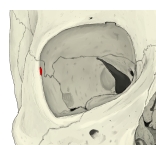 Right orbit (anterior view)
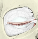 Left eyelids and orbital septum
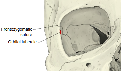
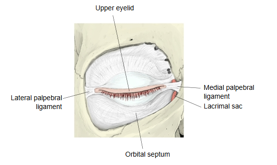
It is a landmark in eyelid and orbital surgery, for example when the lateral corner of the eyelids is reattached. Its size varies; in some skulls it is hardly visible, and some anatomy texts do not list it.
Related terms
- Zygomatic bone
- Orbital margin of zygomatic bone
- Frontozygomatic suture
- Lateral palpebral ligament
- Levator palpebrae superioris
- Orbit
References and sources
- Further reading: Zygomatic bone, Wikipedia.
- Further reading: Lateral palpebral ligament, Wikipedia.
- Federative International Programme for Anatomical Terminology (FIPAT). Terminologia Anatomica, 2nd ed. (TA2), 2019.