Thin ascending limb (Latin: pars ascendens tubuli intermedii)
Also called the thin ascending limb of the loop of Henle
Overview
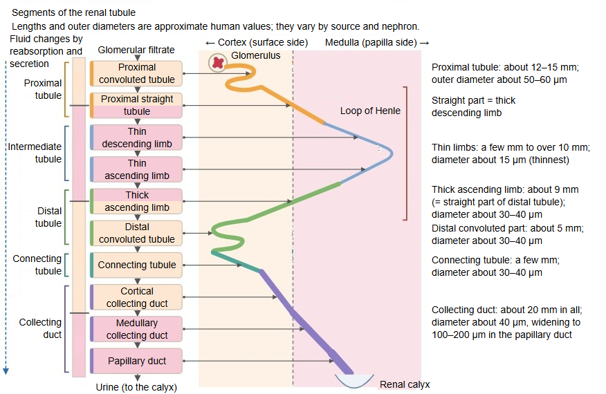
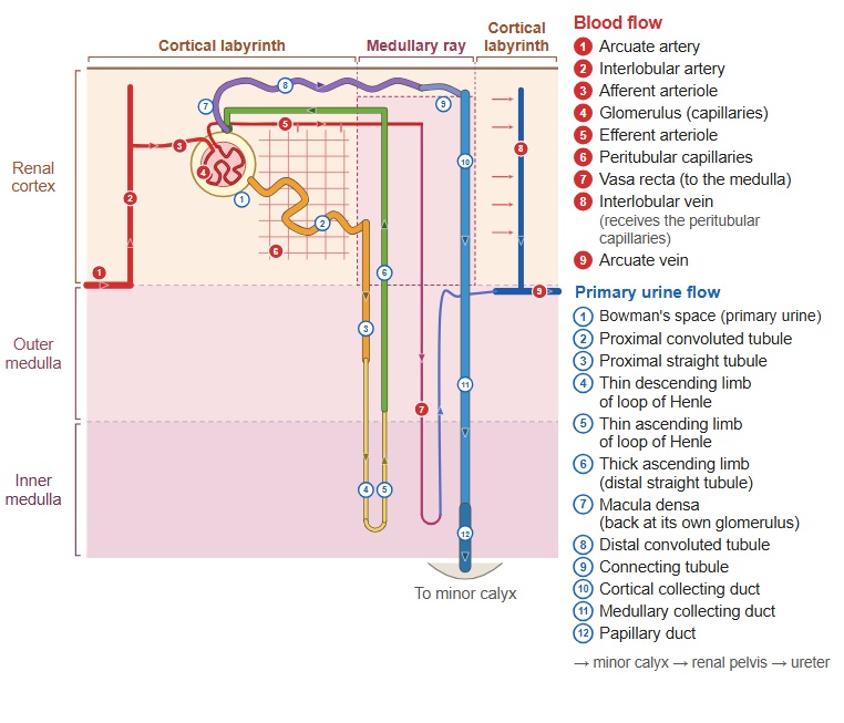
· The thin ascending limb is the thin-walled part of the loop of Henle that follows the bend of the loop, where the thin descending limb turns, and climbs through the renal medulla towards the renal cortex. It continues into the thick ascending limb. Together with the thin descending limb it is called the intermediate tubule.
· Only juxtamedullary nephrons, whose long loops reach deep into the inner zone of the medulla, have a thin ascending limb. In cortical nephrons (short loops) the thin descending limb passes into the thick ascending limb before the bend, so there is no thin ascending limb. The border between the thin and thick ascending limbs lies roughly at the border between the inner and outer zones of the medulla.
· The point where the thin part changes back into the thick part varies: in long loops it lies in the ascending limb, in short loops already in the descending limb.
· Its wall is a simple squamous epithelium, the same as in the thin descending limb; with the light microscope the two limbs are hard to tell apart.
· It is almost impermeable to water but readily lets sodium and chloride ions through. Salt leaves mainly by passive diffusion into the surrounding interstitium, so the fluid in the tubule becomes gradually more dilute. Because salt leaves without water, the inner medulla keeps its high osmotic pressure, which helps the kidney make concentrated urine.
Reference websites
[Wikipedia]
· Loop of Henle, Wikipedia.
[Websites with illustrations]
· Tubules and collecting system (Digital Histology)
· Tubules and collecting system (thin limbs) (Digital Histology)
· Gray's Anatomy, figure 1128 (Wikimedia Commons)

· Transport along the nephron (Wikimedia Commons)

Related terms
- Loop of Henle
- Thin descending limb
- Thick ascending limb
- Intermediate tubule
- Juxtamedullary nephron
- Renal medulla
- Renal tubule
References and sources
- Further reading: Loop of Henle, Wikipedia.
- Federative International Programme for Anatomical Terminology (FIPAT). Terminologia Anatomica, 2nd ed. (TA2), 2019.