Renal medulla (Latin: medulla renalis)
Overview
· The renal medulla is the inner part of the renal parenchyma of the kidney, enclosed by the renal cortex. It consists of several cone-shaped renal pyramids; cortex extends between neighbouring pyramids to form the renal columns.
· The straight parts of the renal tubules (the loops of Henle), the collecting ducts and the vasa recta run side by side, so a cut surface shows fine striations.
· The outer zone of the medulla, next to the cortex, is reddish because it is rich in blood vessels; the inner zone is paler (greyish) and contains mainly collecting ducts and thin tubules. The outer zone is further divided into an outer and an inner stripe.
· The tip of each renal pyramid forms a renal papilla, which projects into a minor calyx.
· The medullary interstitium is kept at a high osmotic pressure by the countercurrent system; this is used to reabsorb water and make concentrated urine.
· The medulla receives only a small part of the renal blood flow and has little oxygen, so it is easily affected by low blood pressure and hypoxia. Drugs such as painkillers (NSAIDs) can cause necrosis of the renal papillae (papillary necrosis).
- Renal cortex
- Renal corpuscle : glomerulus and Bowman's capsule.
- Proximal tubule : takes back water and nutrients from the filtrate.
- Distal tubule : adjusts electrolytes and water.
- Cortical collecting duct : collects urine from several distal tubules.
- Renal medulla
- Renal pyramid : the cone-shaped tissue that forms the medulla; it contains:
- Loop of Henle : part of the countercurrent system that concentrates urine.
- Medullary collecting duct : carries urine to the renal papilla.
- Papillary duct : opens at the tip of the papilla and drains urine into a minor calyx.
- Renal pyramid : the cone-shaped tissue that forms the medulla; it contains:
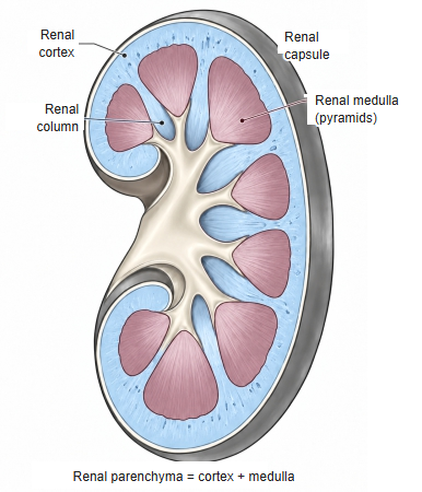
renal medulla (click to enlarge)
Reference websites
[Wikipedia]
· Renal medulla, Wikipedia.
· Kidney, Wikipedia.
[Websites with illustrations]
· The anatomy of the kidney (Interactive Biology)
· Gross anatomy of the kidney (Lumen Learning, Anatomy and Physiology II)
· Kidney anatomy (Blausen) (Wikimedia Commons)

· Hydronephrosis (ultrasound) (NYSORA)
Related terms
- Renal cortex
- Renal pyramid
- Renal papilla
- Renal column
- Loop of Henle
- Medullary collecting duct
- Kidney
References and sources
- Further reading: Renal medulla, Wikipedia.
- Further reading: Kidney, Wikipedia.
- Federative International Programme for Anatomical Terminology (FIPAT). Terminologia Anatomica, 2nd ed. (TA2), 2019.