Intermediate tubule (Latin: tubulus intermedius)
Overview
⇒ Structure of the whole kidney
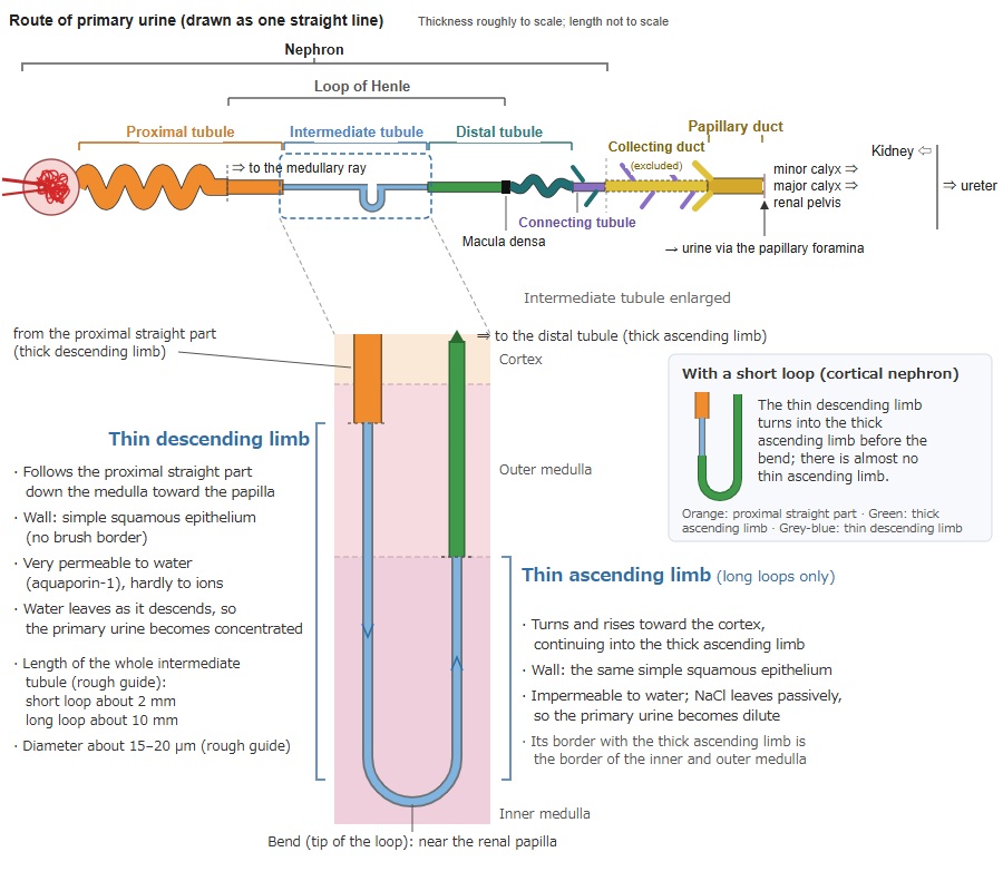
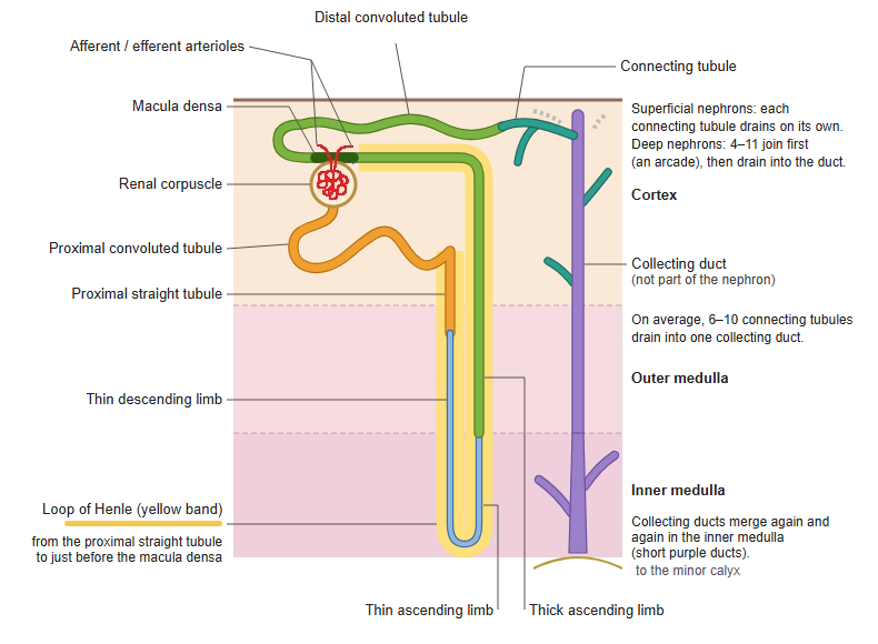
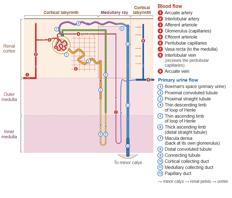
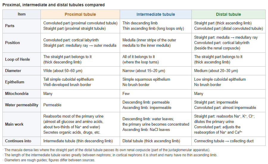
· It is the second part of the renal tubule: it begins at the straight part of the proximal tubule (the proximal straight tubule, thick descending limb) and ends at the straight part of the distal tubule (the thick ascending limb, distal straight tubule).
· In the nephron (renal corpuscle + renal tubule) it lies between the proximal tubule and the distal tubule.
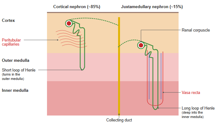
· It is divided into the thin descending limb, before the bend at the tip of the loop of Henle, and the thin ascending limb, after the bend; both lie in the renal medulla. Only juxtamedullary nephrons, with long loops, have a thin ascending limb; in cortical nephrons, with short loops, the thin descending limb passes into the thick ascending limb just before the tip of the loop.
· Older textbooks divide the loop of Henle by its calibre into three parts: first a thick part, then a thin part, and then a thick part again. The thin middle part is the intermediate tubule.
· Its epithelium is a simple squamous epithelium whose cells bulge into the lumen only where the nucleus lies. It has no brush border, and the cells contain few organelles. 'Thin' and 'thick' refer to the height of the epithelial cells, not to the size of the lumen.
· The thin descending limb lets water through easily, while the thin ascending limb is impermeable to water but lets sodium and other salts through. This difference is part of the countercurrent multiplier system of the loop of Henle, which keeps the osmotic pressure of the renal medulla higher the deeper one goes and so helps to make concentrated urine.
· The 'intermediate part' (pars intermedia) of older anatomy textbooks meant the cortical segment that follows the ascending limb (the distal convoluted and connecting tubules). It is not the intermediate tubule described here.
| Item | Thin descending limb | Thin ascending limb |
|---|---|---|
| Position | From the outer medulla down toward the renal papilla (in long loops deep into the inner medulla) | Inner medulla only (rises from the bend of a long loop) |
| Shape | Follows the proximal straight part and runs straight down | Runs straight up after the bend |
| Length | Varies greatly between nephrons (whole intermediate tubule, rough guide): short loops about 2 mm, long loops about 10 mm | |
| Diameter | About 15–20 µm (rough guide; about one third of the proximal tubule) | |
| Epithelium | Simple squamous epithelium, no brush border; the cell bulges into the lumen only at the nucleus | Simple squamous epithelium (hard to tell from the descending limb in the light microscope) |
| Water | Very permeable (aquaporin-1) | Impermeable |
| NaCl | Hardly permeable | Very permeable |
| Main work | Water leaves, so the primary urine inside becomes concentrated (most concentrated at the bend) | NaCl leaves passively, so the primary urine inside becomes dilute (keeps the osmotic pressure of the inner medulla high) |
| Found in | All nephrons | Long loops only (juxtamedullary nephrons) |
| Continues into | Thin ascending limb (in short loops, the thick ascending limb) | Thick ascending limb (distal straight tubule) |
| Loop of Henle | Included | Included |
· In short loops (cortical nephrons) the thin descending limb passes into the thick ascending limb just before the bend, so there is almost no thin ascending limb. 'Thin' and 'thick' refer to the height of the epithelial cells, not to the size of the lumen. Figures are rough guides and differ between sources.
→ Proximal, intermediate and distal tubules compared (table)
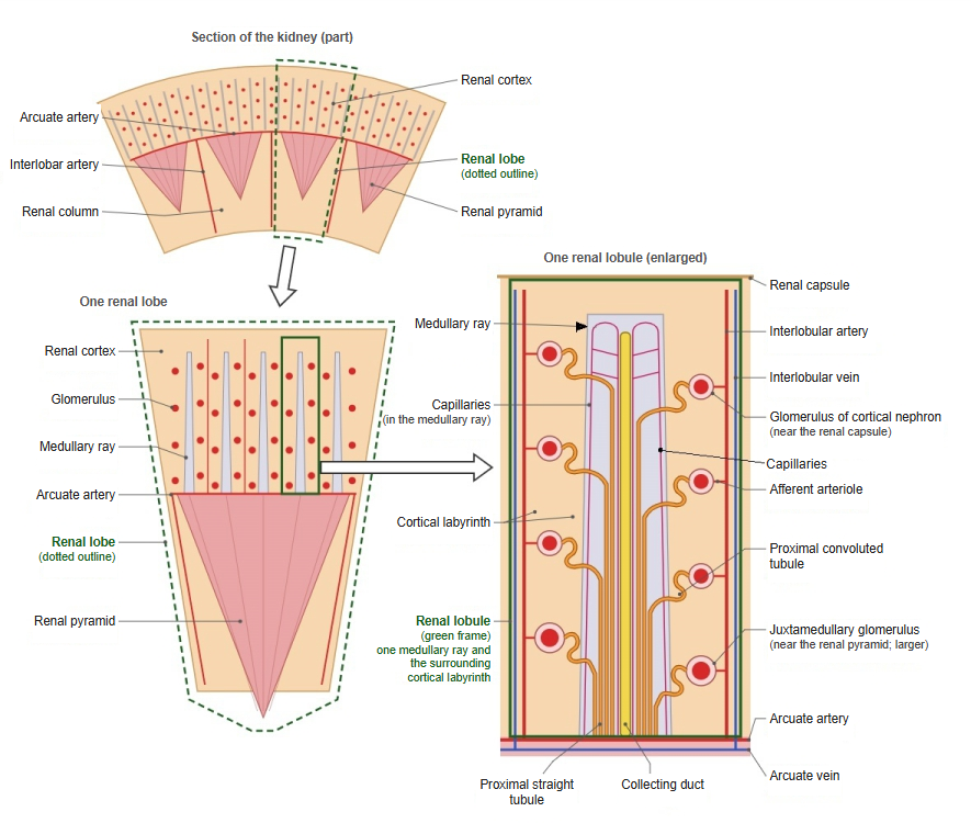
 Renal lobe and renal lobule
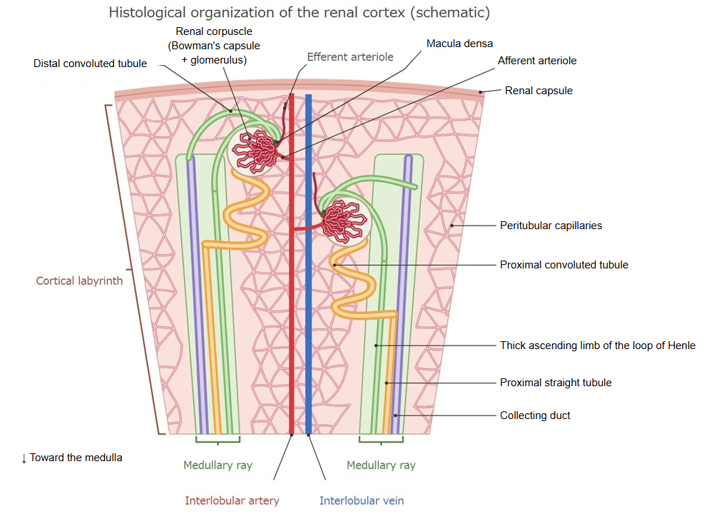
 Tissue organization of the renal cortex
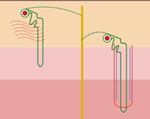 Cortical and juxtamedullary nephrons
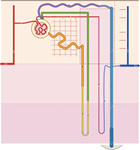 Nephron and blood vessels
Reference websites
[Wikipedia]
· Loop of Henle, Wikipedia.
· Nephron, Wikipedia.
[Websites with illustrations]
· Tubules and collecting system (Digital Histology)
· Tubules and collecting system (2) (Digital Histology)
· Gray's Anatomy, figure 1128 (Wikimedia Commons)

· Nephron transport diagram (Wikimedia Commons)
