Renal cortex (Latin: cortex renalis)
- Overview
- Components
- Comparison of the cortex and medulla
- Reference websites
- (Wikipedia, websites with illustrations)
- Related terms
Overview
· The renal cortex is the outer part of the parenchyma of the kidney, lying just beneath the fibrous capsule. It is about 6–10 mm thick.
· All the nephrons that belong to one medullary ray form a cortical lobule. There is no connective tissue boundary between lobules, so the lobule is a conceptual unit.
· Filtration of blood in the glomeruli takes place in the cortex. The cortex also makes erythropoietin, the hormone that stimulates the production of red blood cells.
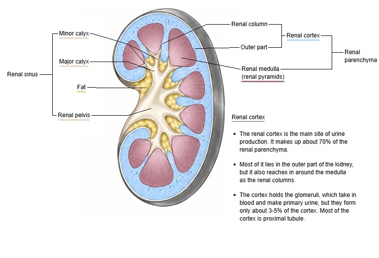
- Renal cortex
- Renal corpuscle : glomerulus and Bowman's capsule
- Proximal tubule : reabsorbs water and nutrients from the primary urine (convoluted part)
- Distal tubule : adjusts salts and water (convoluted part)
- Cortical collecting duct : collects the primary urine from several connecting tubules
- Renal medulla
- Renal pyramid : the cone-shaped tissue that forms the medulla; it contains:
- Loop of Henle : U-shaped part (proximal straight tubule, intermediate tubule, distal straight tubule)
- Medullary collecting duct : carries the primary urine toward the renal papilla
- Papillary duct : opens at the tip of the renal papilla and empties the primary urine into a minor calyx, where it becomes urine
- Renal pyramid : the cone-shaped tissue that forms the medulla; it contains:
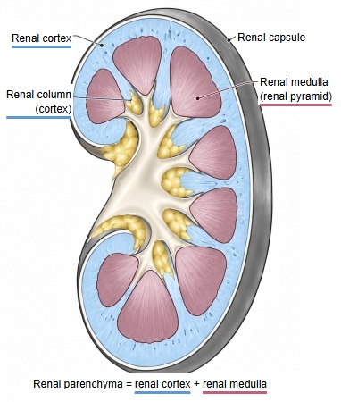
Components
Division by position
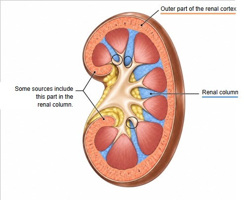
By its position, the renal cortex can be divided into the following two parts.
- Outer part : the part outside the line joining the bases of the renal pyramids (some sources call it the cortical arches, but the term is not in common use)
- Renal column : the part that enters between the renal pyramids
Division by structure
cortex
- Cortical labyrinth : renal corpuscles, proximal convoluted tubules, distal convoluted tubules, connecting tubules, juxtaglomerular apparatus, interlobular arteries and veins, afferent and efferent arterioles, capillariesabout 70–75%
- Medullary rays : proximal straight tubules, thick ascending limbs of the loop of Henle, collecting ducts, capillariesabout 25–30%
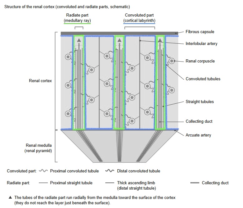
· Renal corpuscle: made up of the glomerulus and Bowman's capsule; it filters the blood to make the primary urine. Renal corpuscles are found only in the cortex.
· Renal tubules: the cortical labyrinth contains the proximal convoluted tubules, distal convoluted tubules and connecting tubules; the proximal straight tubules and the thick ascending limbs (the distal straight tubules) run in the medullary rays.
· Collecting ducts: the cortical collecting ducts descend in the medullary rays and continue into the medulla.
· Juxtaglomerular apparatus: lies at the vascular pole of the renal corpuscle and consists of the macula densa, the juxtaglomerular cells and the extraglomerular mesangial cells (see juxtaglomerular apparatus).
· Blood vessels: the interlobar arteries and veins pass through the renal columns. From the arcuate arteries at the border between the cortex and the medulla, the interlobular arteries ascend through the cortex, followed by the afferent arteriole → glomerular capillaries → efferent arteriole → peritubular capillaries.
cortex
- Cortical labyrinth : the convoluted part; renal corpuscles and convoluted tubules gather here.
- Renal corpuscle : filters the blood to make the primary urine.
- Glomerulus : a tuft of capillaries; the blood is filtered here.
- Bowman's capsule : the sac around the glomerulus; receives the filtered primary urine.
- Proximal convoluted tubule : the coiled part of the tubule that leaves the renal corpuscle (about 48%*).
- Distal convoluted tubule : the coiled part that follows the loop of Henle.
- Renal corpuscle : filters the blood to make the primary urine.
- Medullary ray : the radiate part; runs radially from the medulla toward the surface of the cortex.
- Straight tubule : the straight part of the renal tubule.
- Cortical collecting duct : collects the primary urine from the distal tubules and carries it to the medulla.
- Renal column : cortex that extends between the renal pyramids.
- Blood vessels : interlobular arteries and veins, afferent and efferent arterioles and others.
Note: all the nephrons that belong to one medullary ray form a cortical lobule (there is no boundary membrane; it is a conceptual unit). * Share of the cortical volume taken by the proximal convoluted tubule (a value measured in the rat); no reliable figures are given for the other parts.
Comparison of the cortex and medulla
· The table below compares the components of the cortex and the medulla in brief.
| Item | Renal cortex | Renal medulla: outer zone | Renal medulla: inner zone |
|---|---|---|---|
| Share of the renal parenchyma | about 70% (60–80% depending on the source) | about 30% (20–40% depending on the source) | |
| Components | |||
| Divisions | cortical labyrinth, medullary rays, renal columns | outer zone of the renal pyramids (divided into an outer and an inner stripe) | inner zone of the renal pyramids (its tip is the renal papilla) |
| Renal corpuscle | renal corpuscle (glomerulus and Bowman's capsule) | none | |
| Renal tubules | proximal convoluted tubule distal convoluted tubule connecting tubule (in the medullary rays: proximal straight tubule and thick ascending limb) | proximal straight tubule thin descending limb thick ascending limb (= distal straight tubule) | thin descending limb thin ascending limb |
| Collecting ducts | cortical collecting duct | medullary collecting duct | medullary collecting duct, papillary duct |
| Juxtaglomerular apparatus | macula densa, juxtaglomerular cells, extraglomerular mesangial cells | none | |
| Blood vessels | interlobar arteries and veins (in the renal columns) interlobular arteries and veins afferent and efferent arterioles glomerular capillaries peritubular capillaries | vasa recta (descending and ascending vasa recta) | |
| Contents by volume (approximate) | |||
| Renal tubules and collecting ducts (including the fluid inside them) | about 75–80% | about 65–70% | about 30–55% |
| Blood (inside the vessels) | about 10% | about 25% | about 35% |
| Interstitium | about 7–9% | about 3–10% | about 10–40% |
| Glomeruli | about 3–5% | none | none |
Note: The arcuate arteries and veins run along the border between the cortex and the medulla.
Note: The medullary rays lie within the cortex, but like the medulla they contain straight renal tubules and collecting ducts running side by side.
Note: The contents are approximate values measured mainly in the rat (the glomerular value comes from measurements in humans). Renal tubules, collecting ducts and others are the value left after subtracting the other parts from 100%.
Note: The deeper part of the medulla has more blood and interstitium, so the share of the renal tubules and collecting ducts becomes smaller.
| Component | Cortex | Outer medulla | Inner medulla |
|---|---|---|---|
| ■ Renal tubules, collecting ducts and others (including the fluid inside them) | about 75–80% | about 65–70% | about 30–55% |
| ■ Blood (inside the vessels) | about 10% | about 25% | about 35% |
| ■ Interstitium | about 7–9% | about 3–10% outer stripe about 3–5% inner stripe about 10% | about 10–40% at the border about 10% toward the papillary tip 30–40% |
| ■ Glomeruli | about 3–5% | none | none |
Note: Approximate values, mainly measured in the rat. Renal tubules, collecting ducts and others are the value left after subtracting the other parts from 100%.
Note: Blood means the blood inside the vessels, not the vessel walls. The glomerular value comes from measurements in humans (about 4 cm3 in total in one kidney).
Note: Deeper in the medulla there is more blood and interstitium, so the share of the renal tubules and collecting ducts becomes smaller.
Reference websites
[Wikipedia]
· Renal cortex, Wikipedia.
· Kidney, Wikipedia.
[Websites with illustrations]
· The anatomy of the kidney (Interactive Biology)
· Gross anatomy of the kidney (Lumen Learning, Anatomy and Physiology II)
· Kidney cross-section (EBM Consult)
· Kidney anatomy (Blausen) (Wikimedia Commons)

Related terms
- Renal medulla
- Renal column
- Renal corpuscle
- Cortical labyrinth
- Medullary rays
- Fibrous capsule of kidney
- Nephron
References and sources
- Further reading: Renal cortex, Wikipedia.
- Further reading: Kidney, Wikipedia.
- Federative International Programme for Anatomical Terminology (FIPAT). Terminologia Anatomica, 2nd ed. (TA2), 2019.