Glomerular filtration rate
Also called GFR
Overview
· The glomerular filtration rate (GFR) is the volume of fluid (primary urine) filtered from the blood into the capsular space (Bowman's space) by all the glomeruli of both kidneys in a given time, usually one minute. It is regarded as the most important measure of how well the kidneys work.
· In a healthy adult it is about 100-125 mL per minute, or about 150-180 L per day. Because it varies with age, sex and body size, it is usually expressed per 1.73 m² of body surface area. It falls gradually with age.
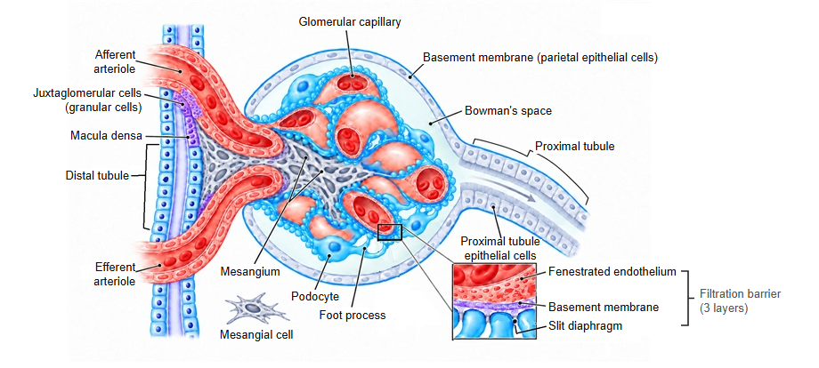
· GFR is set by the difference between the blood pressure in the glomerular capillaries, which drives filtration, and the opposing colloid osmotic pressure of the plasma proteins and pressure in the capsular space, together with the area and permeability of the filtration barrier (endothelial cells, glomerular basement membrane and podocytes). Glomerular pressure is adjusted by the calibre of the afferent and efferent arterioles; the efferent arteriole leaving the glomerulus is narrower than the afferent one, and this difference helps build up the filtration pressure.
· Even when systemic blood pressure changes, autoregulation of the arterioles and feedback through the juxtaglomerular apparatus (including the macula densa), called tubuloglomerular feedback, keep GFR nearly constant over a certain range.
· Clinically it is measured from the clearance of inulin or creatinine into the urine, and more often estimated (eGFR) from the blood level of creatinine (or cystatin C) together with age and sex. A GFR below 60 mL/min per 1.73 m² that lasts for three months or more indicates chronic kidney disease (CKD).
Reference websites
[Wikipedia]
· Glomerular filtration rate, Wikipedia.
· Renal function, Wikipedia.
[Websites with illustrations]
· Physiology of urine formation (OpenStax Anatomy and Physiology 2e)
· Physiology of the nephron (diagram) (Wikimedia Commons)

· Estimated glomerular filtration rate (eGFR) (National Kidney Foundation)
· Glomerular filtration rate (NIDDK (NIH))
Related terms
- Glomerulus
- Primary urine
- Capsular space
- Glomerular capillary
- Glomerular basement membrane
- Juxtaglomerular apparatus
- Macula densa
References and sources
- Further reading: Glomerular filtration rate, Wikipedia.
- Further reading: Renal function, Wikipedia.
- Federative International Programme for Anatomical Terminology (FIPAT). Terminologia Anatomica, 2nd ed. (TA2), 2019.