Podocyte (Latin: podocytus)
Also called the visceral epithelial cell
Overview
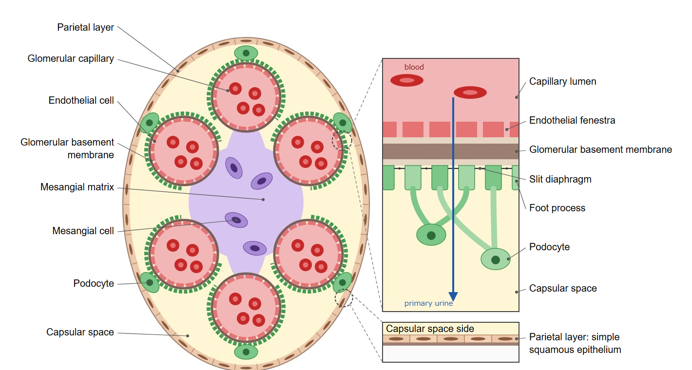
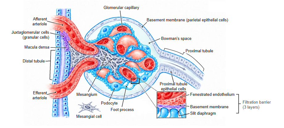
· A podocyte is a specialized epithelial cell that forms the visceral layer of the glomerular capsule (Bowman's capsule). Its processes wrap around the glomerular capillaries like the arms of an octopus. It is also called a visceral epithelial cell.
· Thick primary processes arise from the cell body and divide into many foot processes (pedicels). The foot processes of neighbouring podocytes interlock, and the narrow gaps between them are called filtration slits.
· Each filtration slit is bridged by a thin membrane, the slit diaphragm. The slit diaphragm contains the protein nephrin and acts as a sieve: water and small molecules pass, but proteins are held back.
· The foot processes are attached to the outer surface of the glomerular basement membrane. Podocytes also help to build and maintain this basement membrane.
· The filtrate that passes the filtration slits enters the capsular space. Podocytes are the last barrier of the filtration barrier, which consists of the endothelial pores (fenestrations), the basement membrane and the slit diaphragm.
· Mutations of nephrin and other genes cause congenital nephrotic syndrome. When podocytes are injured, their foot processes flatten out (foot process effacement) and protein appears in the urine (proteinuria).
Reference websites
[Wikipedia]
· Podocyte, Wikipedia.
· Bowman's capsule, Wikipedia.
· Glomerulus (kidney), Wikipedia.
[Websites with illustrations]
· Renal corpuscle (diagram) (Wikimedia Commons)

· Filtration barrier (diagram) (Wikimedia Commons)

· Renal corpuscle (electron micrograph) (Histology Guide)
· Renal corpuscle (Digital Histology)
Related terms
- Parietal layer of glomerular capsule
- Vascular pole
- Urinary pole
- Visceral layer of glomerular capsule
- Glomerular basement membrane
- Glomerular capillary
- Capsular space
- Mesangium
- Kidney
References and sources
- Further reading: Podocyte, Wikipedia.
- Further reading: Bowman's capsule, Wikipedia.
- Further reading: Glomerulus (kidney), Wikipedia.
- Federative International Programme for Anatomical Terminology (FIPAT). Terminologia Anatomica, 2nd ed. (TA2), 2019.