Capsular space
Also called Bowman's space or the urinary space
Overview
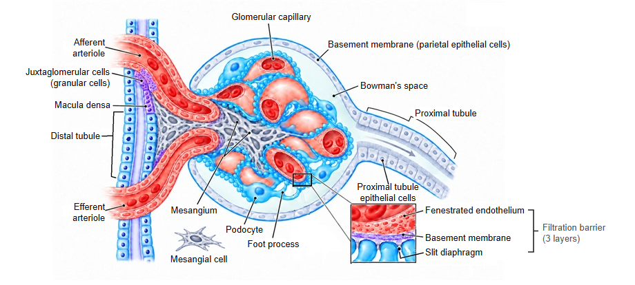
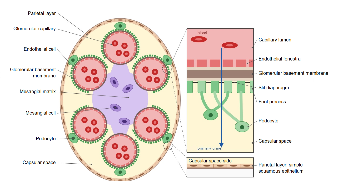
· The capsular space (Bowman's space) is the narrow cavity inside the glomerular capsule, between its visceral layer and parietal layer. The fluid filtered from the glomerulus (the primary urine) enters it first. It is also called the urinary space.
· Its inner wall is formed by the podocytes (the visceral layer) that cover the glomerular capillaries; its outer wall is the simple squamous epithelium of the parietal layer.
· Blood is filtered into the capsular space through the endothelium of the glomerular capillaries, the glomerular basement membrane and the filtration slits between the podocyte foot processes. Small molecules such as water, ions, glucose and amino acids pass through; blood cells and large proteins do not.
· At the urinary pole the capsular space opens into the lumen of the proximal tubule, so the filtrate flows straight on into the renal tubule.
· The volume of fluid filtered by the glomeruli per minute, the glomerular filtration rate (GFR), is the standard measure of kidney function; a low GFR is a sign of kidney failure.
Reference websites
[Wikipedia]
· Bowman's capsule, Wikipedia.
· Renal corpuscle, Wikipedia.
[Websites with illustrations]
· Renal corpuscle (Digital Histology)
· Renal corpuscle (micrograph) (SIU School of Medicine histology)
· Renal corpuscle (diagram) (Wikimedia Commons)

· Glomeruli histology (kidneypathology.com)
Related terms
- Glomerular capillary
- Mesangium
- Vascular pole
- Visceral layer of glomerular capsule
- Parietal layer of glomerular capsule
- Podocyte
- Urinary pole
- Proximal tubule
- Glomerular basement membrane
- Kidney
References and sources
- Further reading: Bowman's capsule, Wikipedia.
- Further reading: Renal corpuscle, Wikipedia.
- Federative International Programme for Anatomical Terminology (FIPAT). Terminologia Anatomica, 2nd ed. (TA2), 2019.