Fourth ventricle (Latin: ventriculus quartus)
There are three kinds of ventricles and four ventricles in all, because there is a lateral ventricle on each side.
- Lateral ventricles : one in each cerebral hemisphere: left and right lateral ventricles.
- Third ventricle : lies in the diencephalon.
- Fourth ventricle : lies between the medulla oblongata and pons and the cerebellum.
· Median aperture of the fourth ventricle (foramen of Magendie): a single opening in the midline, at the lower part of the roof.
· Lateral apertures of the fourth ventricle (foramina of Luschka): one on each side, opening at the outer end of the left and right lateral recesses.
The inner surface of the fourth ventricle is covered by ependyma. It is divided into the floor, the rhomboid fossa, and the roof above it, the roof of the fourth ventricle, formed by the superior and inferior medullary vela.
ventricle
- Rhomboid fossa : the shallow, diamond-shaped fossa that forms the floor of the fourth ventricle.
- Roof of fourth ventricle : the roof of the fourth ventricle, the part covered by the cerebellum.
The choroid plexus of the fourth ventricle is paired. The two strips begin at the lower end of the ventricle, run upwards side by side to the inferior medullary velum, and there turn outwards towards the lateral recesses.
Parts of the choroid tissue of the fourth ventricle protrude through the median and lateral apertures and lie outside the ventricle.
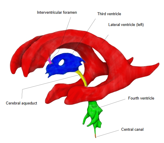 Ventricles: left side (coloured)
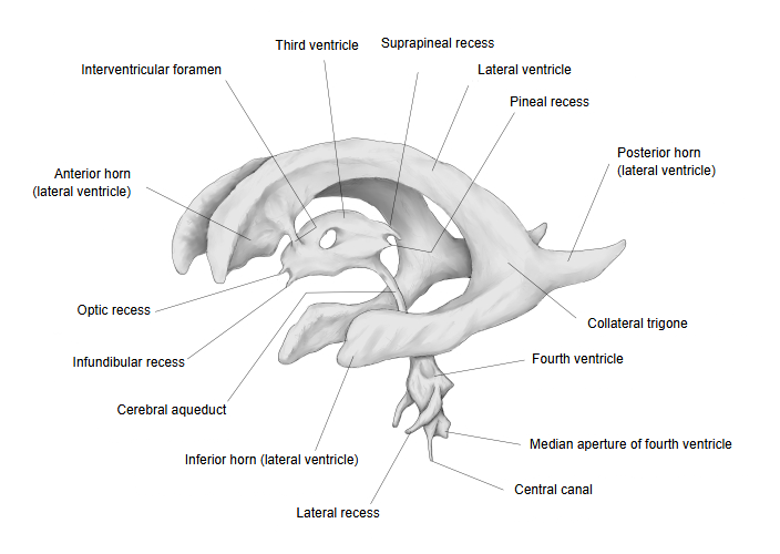 Ventricles: left side
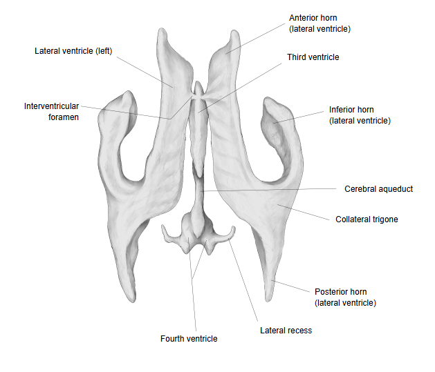
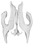 Ventricles: from above
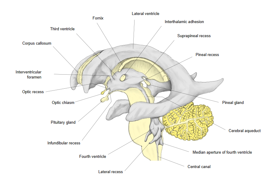 Ventricles in the brain (midline view)
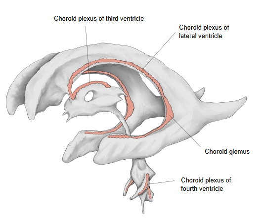 Choroid plexuses
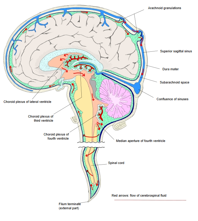
 Flow of cerebrospinal fluid
Reference websites
[Wikipedia]
· Fourth ventricle, Wikipedia.
· Ventricular system, Wikipedia.
[Websites with illustrations]
· Floor of the fourth ventricle and the corpora quadrigemina (Gray's Anatomy, fig. 709) (Wikimedia Commons)

Related terms
- Cerebral ventricles
- Third ventricle
- Lateral ventricles
- Rhomboid fossa
- Roof of fourth ventricle
- Choroid plexus of fourth ventricle
- Subarachnoid space
References and sources
- Further reading: Fourth ventricle, Wikipedia.
- Further reading: Ventricular system, Wikipedia.
- Federative International Programme for Anatomical Terminology (FIPAT). Terminologia Anatomica, 2nd ed. (TA2), 2019.