Skip to the content Skip to the Navigation
Visual Anatomy

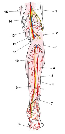
Nerves of the leg (posterior view)

  1. Home
  2. Nerves
  3. Multi-nerve illustrations
  4. Nerves of the leg (posterior view)

⚠ Report an error

← Multi-nerve illustrations: Spinal nerves
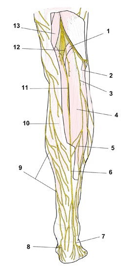
Nerves of the leg (posterior view): numbered illustration
No.NamePage
1PlantarisDetail
2Common fibular nerveDetail
3Lateral sural cutaneous nerveDetail
4Gastrocnemius, lateral headDetail
5Sural communicating branch (common fibular nerve)—
6Sural nerveDetail
7Lateral dorsal cutaneous nerve (sural nerve)—
8Medial calcaneal branches (tibial nerve)—
9Saphenous nerveDetail
10Deep fascia of leg—
11Medial sural cutaneous nerveDetail
12Tibial nerveDetail
13SemimembranosusDetail

More illustrations: Spinal nerves

  • Typical spinal nerve: anterior and posterior rami (schematic)Typical spinal nerve: anterior and posterior rami (schematic)
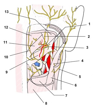
  • Cervical nerves, suboccipital region (posterior view)Cervical nerves, suboccipital region (posterior view)
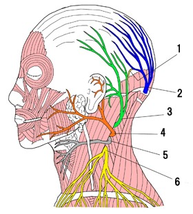
  • Cervical plexus, cutaneous branches (lateral view)Cervical plexus, cutaneous branches (lateral view)
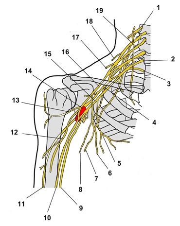
  • Cervical and brachial plexuses, right side (anterior view)Cervical and brachial plexuses, right side (anterior view)
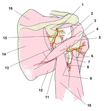
  • Scapular region, nerves and arteries (posterior view)Scapular region, nerves and arteries (posterior view)
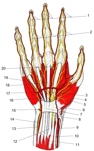
  • Main nerves and arteries of the palm (right hand)Main nerves and arteries of the palm (right hand)
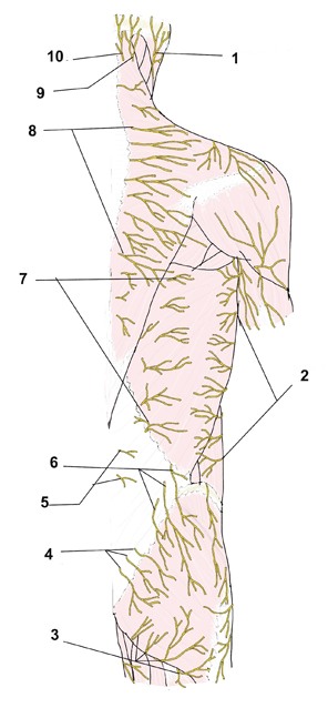
  • Nerves of the back, cutaneous branches (posterior view)Nerves of the back, cutaneous branches (posterior view)
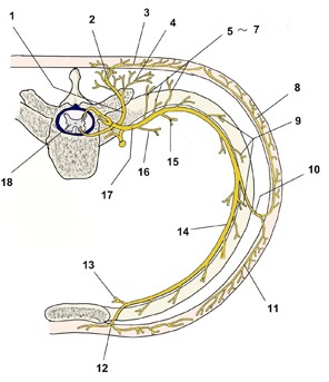
  • Thoracic spinal nerve and intercostal nerve branches (transverse schematic)Thoracic spinal nerve and intercostal nerve branches (transverse schematic)
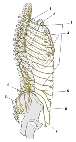
  • Intercostal nerves and nerves of the trunk wall (lateral view)Intercostal nerves and nerves of the trunk wall (lateral view)
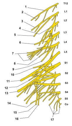
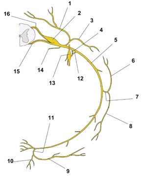
  • Lumbosacral plexus (schematic)Lumbosacral plexus (schematic)
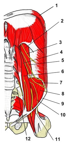
  • Lumbar plexus on the posterior abdominal wall (anterior view)Lumbar plexus on the posterior abdominal wall (anterior view)
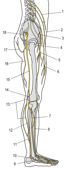
  • Lumbar and sacral plexuses, right lower limb (lateral view)Lumbar and sacral plexuses, right lower limb (lateral view)
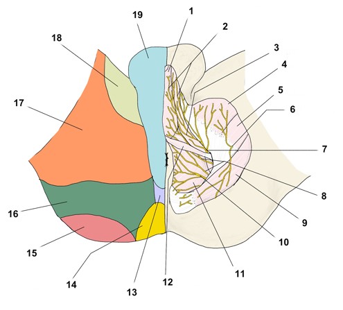
  • Pudendal plexus: nerves and cutaneous territories of the male perineum (inferior view)Pudendal plexus: nerves and cutaneous territories of the male perineum (inferior view)
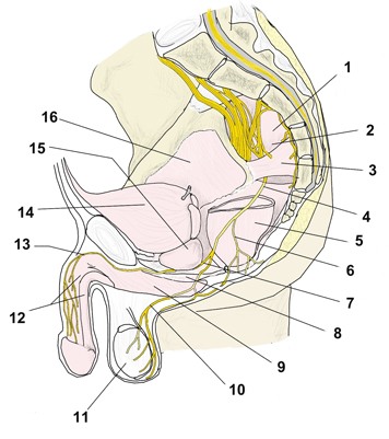
  • Pudendal plexus (median section)Pudendal plexus (median section)
  • Leg, nerves and arteries (posterior view)Leg, nerves and arteries (posterior view)
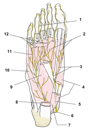
  • Nerves of the sole of the footNerves of the sole of the foot
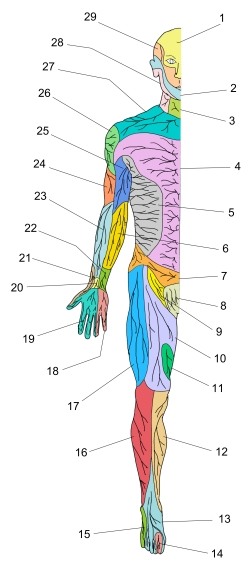
  • Cutaneous nerve distribution (anterior view)Cutaneous nerve distribution (anterior view)
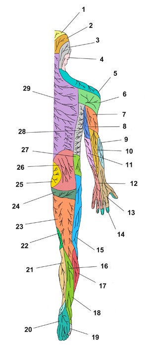
  • Cutaneous nerve distribution (posterior view)Cutaneous nerve distribution (posterior view)

⚠ Report an error

Menu

  • Introduction
    • Regions of the human body
    • Body regions (A–Z)
    • Directional terms
    • List of organs
  • Muscles
    • Name Index
      • SkeletalAlphabetical order
      • SkeletalBy region
      • SmoothList of smooth muscles
    • Illustration List
      • SkeletalSingle-muscle illustrations
      • SkeletalMulti-muscle illustrations
  • Bones
    • Single Bone Details
      • Alphabetical order
      • By region
      • By shape
    • Illustration List
      • Single bones and regions
  • Joints
    • Alphabetical order
    • By region
    • Illustrations
  • Ligaments
    • Alphabetical order
    • By region
  • Nerves
    • Single Nerve Details
      • Alphabetical order
      • By region
    • Illustration List
      • Nerve illustrations
  • Circulatory system
    • Arteries (A–Z)
    • Veins (A–Z)
    • By region
  • Cross-sections
    • By region
  • Splanchnology
    • Alphabetical order
    • By system
  • Sensory Organs
    • Alphabetical order
    • By system
  • Glossary
    • Alphabetical order

Copyright © Visual Anatomy All Rights Reserved.

Powered by WordPress & Lightning Theme by Vektor,Inc. technology.

MENU