Latin: os naviculare · a tarsal bone of the foot
Overview
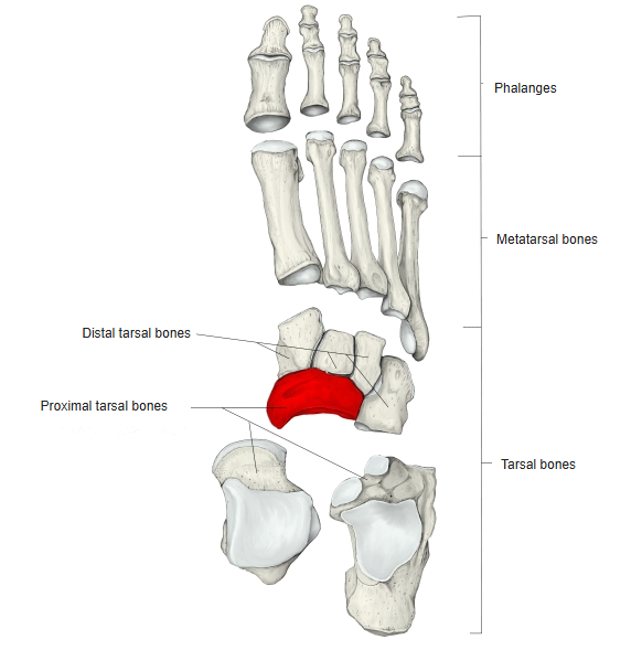
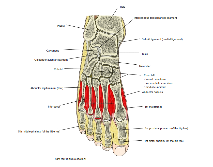
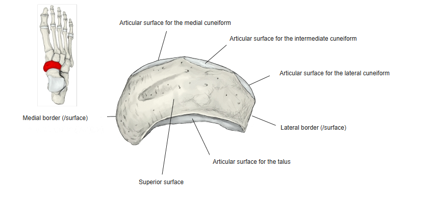
- One of the seven tarsal bones, on the medial side of the foot between the talus behind and the three cuneiforms in front.
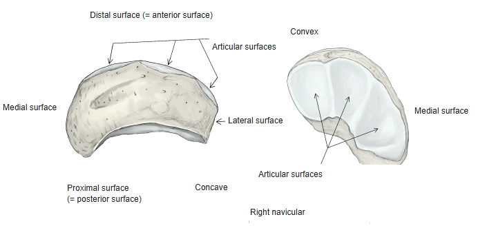
- Flattened from front to back and, as its name says, shaped like a small boat (or cradle).
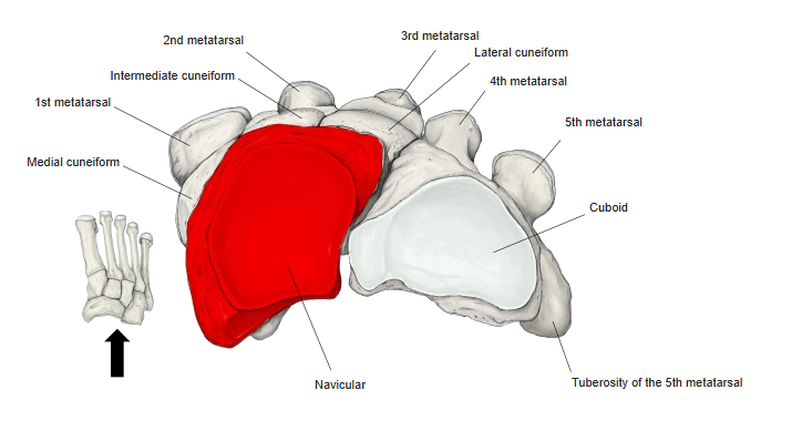
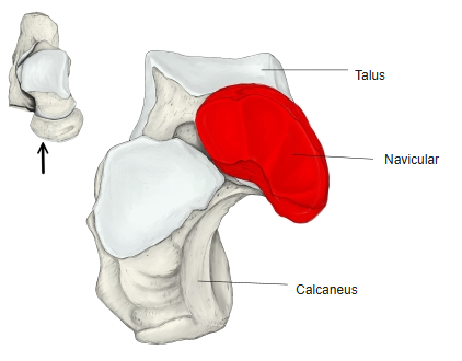
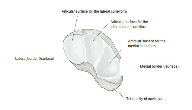
- The proximal (posterior) surface is concave, with one articular surface for the talus; the distal (anterior) surface is convex, with three articular surfaces for the cuneiforms.
- The rough medial surface projects downwards as the tuberosity of the navicular, which can be felt under the skin.
- The lateral surface often articulates with the cuboid; on the plantar surface, near the tuberosity, a shallow oblique groove carries deep fibres of the tibialis posterior tendon.
- Name: from Latin navicula, the diminutive of navis (“ship”) — a “little boat”.
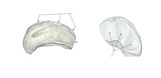
Shape of the navicular
- Bones of the lower limb (one side)
- Pelvic girdle
- Hip bone ← ilium, ischium and pubis fused
- Free lower limb
- Thigh and knee
- Femur
- Patella
- Leg
- Tibia ← big-toe side
- Fibula ← little-toe side
- Foot
- Tarsal bones (7)
- Metatarsal bones (5)
- Phalanges (14)
- Thigh and knee
- Pelvic girdle

Illustrations
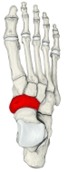
Right foot (dorsal) 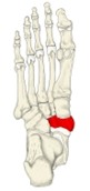
Right foot (plantar) 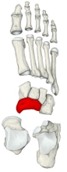
Bones of the foot (separated) 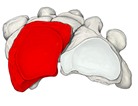
Navicular and distal bones (posterior) 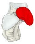
Proximal tarsals (anterior) 
Right foot (section) 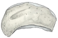
Right navicular (from above) 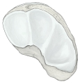
Right navicular (anterior)
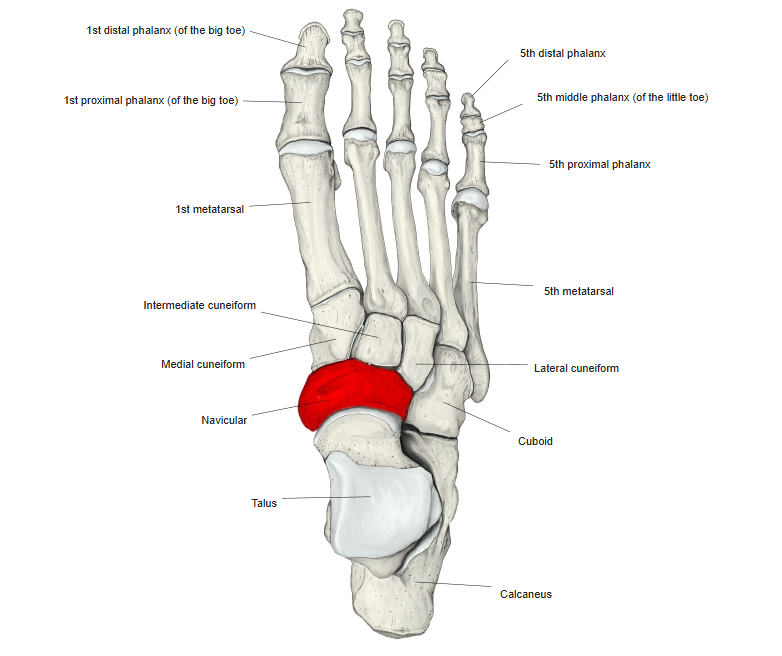
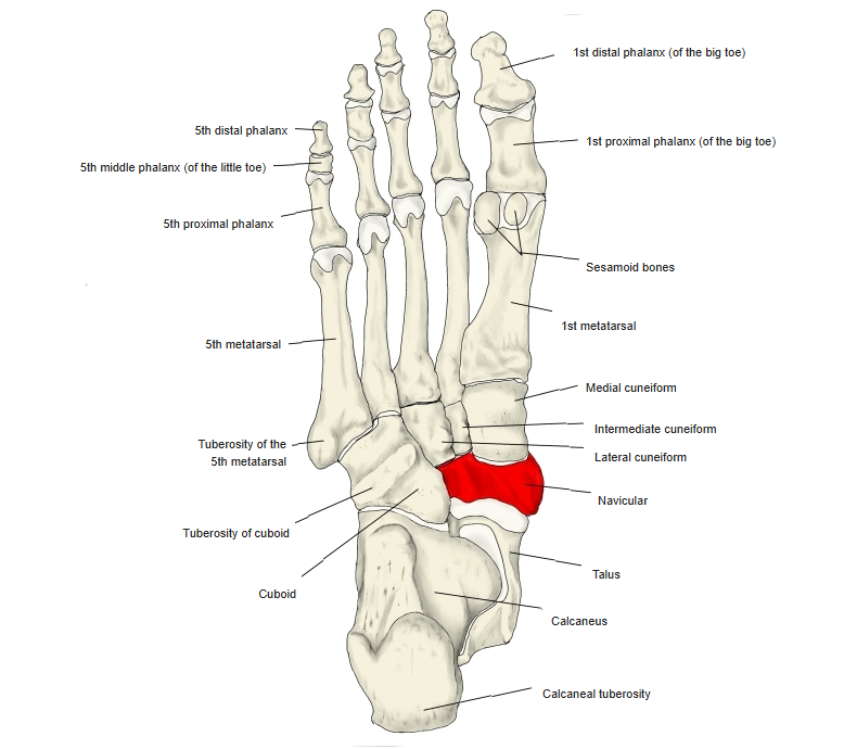
Articulations
| # | Bone | Joint |
|---|---|---|
| 1 | Medial cuneiform | Cuneonavicular joint |
| 2 | Intermediate cuneiform | |
| 3 | Lateral cuneiform | |
| 4 | Talus | Talonavicular (talocalcaneonavicular) joint |
| 5 | Cuboid | Cuboideonavicular joint |
Joints of the navicular
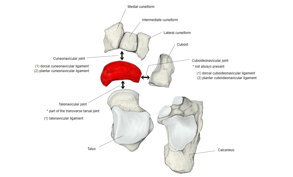
The cuboideonavicular joint is not always present (the two bones are often joined only by ligaments).
Ligaments
The following ligaments attach to the navicular. The list is intended to be comprehensive but may omit minor ligaments.
| # | Ligament | Connects |
|---|---|---|
| 1 | Dorsal cuneonavicular ligaments | Cuneiforms – navicular |
| 2 | Plantar cuneonavicular ligaments | Cuneiforms – navicular |
| 3 | Dorsal talonavicular ligament | Talus – navicular |
| 4 | Dorsal cuboideonavicular ligament | Cuboid – navicular |
| 5 | Plantar cuboideonavicular ligament | Cuboid – navicular |
Muscle attachments
- Origin of: none
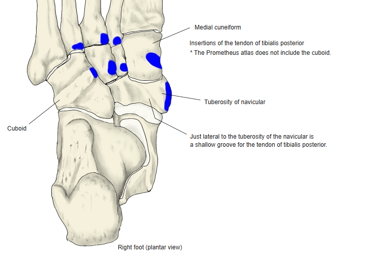
- Insertion of: Tibialis posterior
Insertion: tibialis posterior — tuberosity of the navicular (with slips to the other tarsal bones and metatarsals).
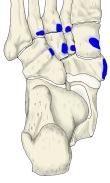
Tibialis posterior insertion
References and sources
- Further reading: Navicular bone, Wikipedia.