Latin: talus · also called the ankle bone
Overview
- One of the proximal tarsal bones and the highest bone of the foot.
- It links the leg bones (tibia and fibula) to the other tarsal bones.
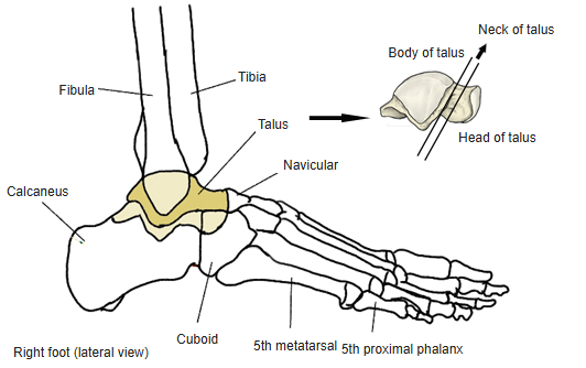
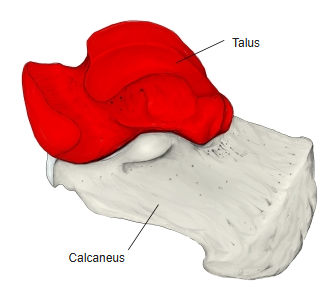
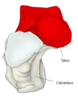
- It has three parts: the body behind, the head projecting forwards and medially, and the neck between them.
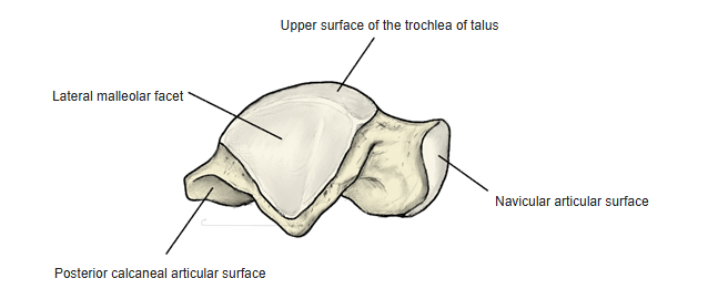
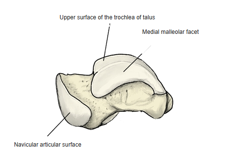
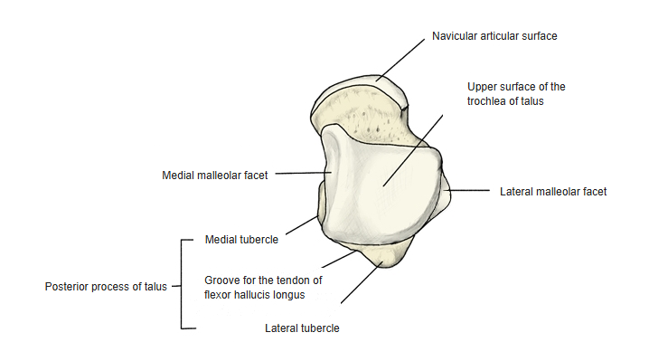
- Body: the trochlea above (superior surface, medial and lateral malleolar facets) for the ankle joint; the posterior process with medial and lateral tubercles and the groove for the flexor hallucis longus tendon between them; the lateral process.
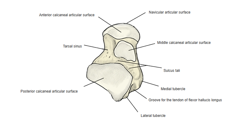
- Underside: the posterior, middle and anterior calcaneal articular surfaces, with the sulcus tali (which forms the tarsal sinus with the calcaneus).
- Head: the navicular articular surface in front.
- No muscles attach to the talus.
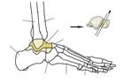
Parts of the talus
- Bones of the lower limb (one side)
- Pelvic girdle
- Hip bone ← ilium, ischium and pubis fused
- Free lower limb
- Thigh and knee
- Femur
- Patella
- Leg
- Tibia ← big-toe side
- Fibula ← little-toe side
- Foot
- Tarsal bones (7)
- Metatarsal bones (5)
- Phalanges (14)
- Thigh and knee
- Pelvic girdle

Illustrations
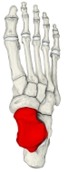
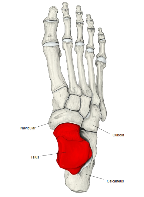
Right foot (dorsal) 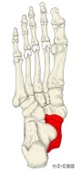
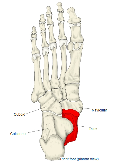
Right foot (plantar) 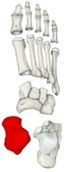
Bones of the foot (separated) 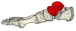
Right foot (medial) 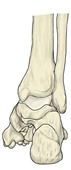
Right ankle (posterior) 
Right foot (section) 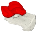
Talus and calcaneus (medial) 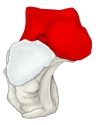
Talus and calcaneus (anterior) 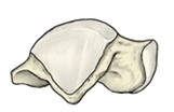
Right talus (lateral) 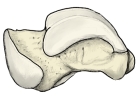
Right talus (medial) 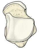
Right talus (from above) 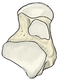
Right talus (from below)
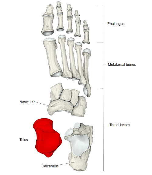
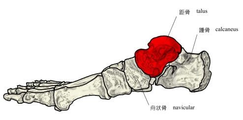
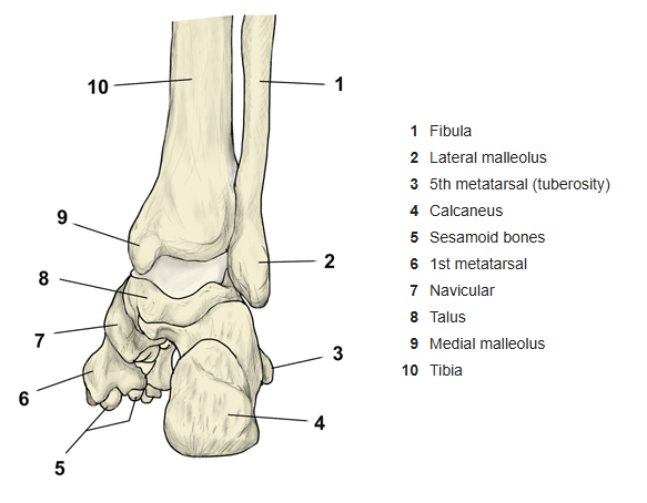
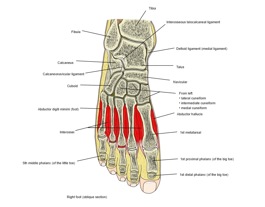
Articulations
| # | Bone | Joint |
|---|---|---|
| 1 | Tibia | Talocrural (ankle) joint |
| 2 | Fibula | |
| 3 | Calcaneus | Subtalar joint |
| 4 | Calcaneus | Talocalcaneonavicular joint |
| 5 | Navicular |
Talocrural joint: trochlea of the talus (superior surface and medial malleolar facet) ↔ inferior articular surface and medial malleolus of the tibia; lateral malleolar facet ↔ lateral malleolus of the fibula. Subtalar joint: posterior calcaneal articular surface ↔ posterior talar articular surface of the calcaneus. Talocalcaneonavicular joint: navicular articular surface and anterior and middle calcaneal articular surfaces ↔ navicular and the anterior and middle talar articular surfaces of the calcaneus.
Ligaments
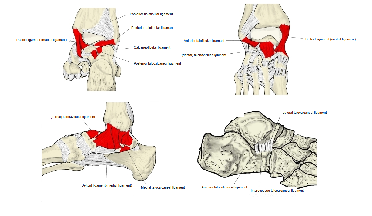
The following ligaments attach to the talus. The list is intended to be comprehensive but may omit minor ligaments.
| # | Ligament | Connects |
|---|---|---|
| 1 | Anterior talofibular ligament | Neck of talus – lateral malleolus of fibula |
| 2 | (Dorsal) talonavicular ligament | Neck of talus – navicular |
| 3 | Posterior talofibular ligament | Posterior process – malleolar fossa of fibula |
| 4 | Posterior talocalcaneal ligament | Posterior process – calcaneus |
| 5 | Deltoid (medial) ligament | Neck and posterior process of talus (also navicular and calcaneus) – medial malleolus of tibia |
| 6 | Medial talocalcaneal ligament | Posterior process (medial tubercle) – sustentaculum tali of calcaneus |
| 7 | Lateral talocalcaneal ligament | Lateral process – calcaneus |
| 8 | Anterior talocalcaneal ligament | Neck of talus – calcaneus |
| 9 | Interosseous talocalcaneal ligament | Sulcus tali – calcaneal sulcus |
Ligaments of the talus
Muscle attachments
- Origin of: none
- Insertion of: none
No muscles attach to the talus.
References and sources
- Further reading: Talus bone, Wikipedia.