Latin: os occipitale
Overview
- The bone forming the back and part of the base of the skull; it surrounds the foramen magnum.
- It has four parts around the foramen magnum: the occipital squama (behind), the two lateral parts (with the occipital condyles) and the basilar part (in front).
- It meets three kinds of bone: the parietal bones, the temporal bones and the sphenoid; with its condyles it also articulates with the atlas.
- Bones of the skull
- Neurocranium (cranial bones)
- Frontal bone
- Parietal bone (2)
- Occipital bone
- Temporal bone (2)
- Sphenoid bone
- Ethmoid bone
- Viscerocranium (facial bones)
- Nasal bone (2)
- Vomer
- Lacrimal bone (2)
- Inferior nasal concha (2)
- Maxilla (2)
- Zygomatic bone (2)
- Palatine bone (2)
- Mandible
- Hyoid bone
- Neurocranium (cranial bones)
Illustrations
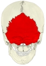
Skull (posterior) 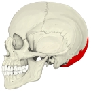
Skull (left lateral) 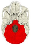
External cranial base 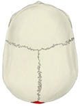
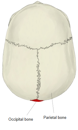
Skull (from above) 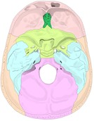
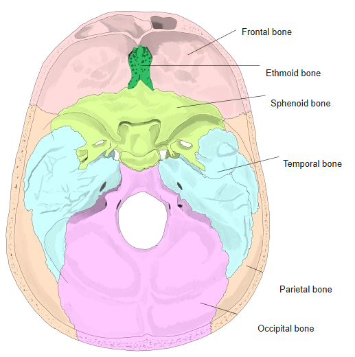
Internal cranial base 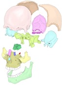
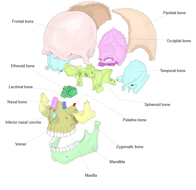
Bones of the skull (separated) 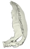
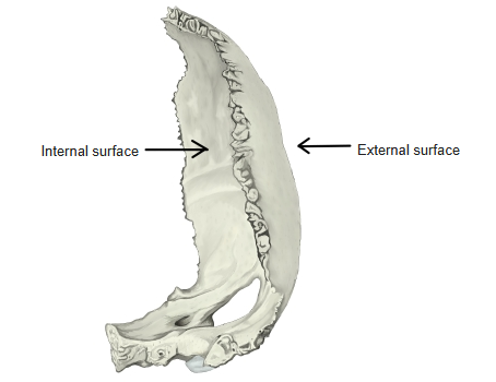
Surfaces of the occipital bone 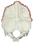
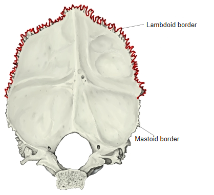
Borders of the occipital squama 
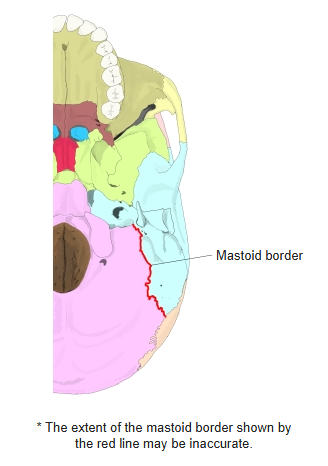
Mastoid border
Articulations
| # | Bone | Joint |
|---|---|---|
| 1 | Parietal bones | Lambdoid suture |
| 2 | Temporal bones | Occipitomastoid suture, petro-occipital fissure |
| 3 | Sphenoid | Spheno-occipital synchondrosis |
| 4 | Atlas (superior articular facets) | Atlanto-occipital joint |
Neighbouring bones (median section) 
Neighbouring bones (posterior) 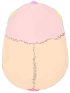
Neighbouring bones (from above) 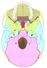
Neighbouring bones (from below)
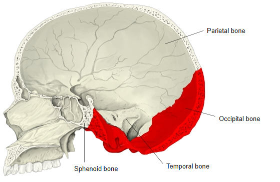
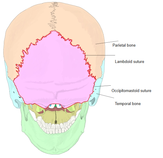
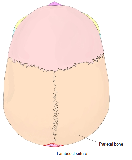
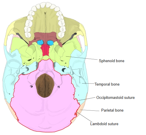
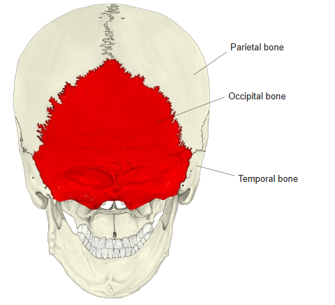
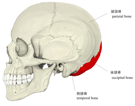
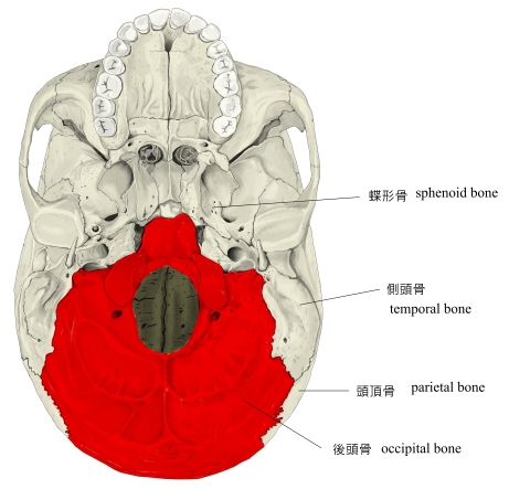
In these figures the occipital bone is outlined in red.
Ligaments
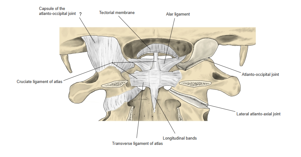
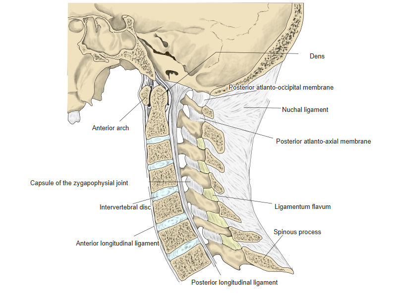
The following ligaments attach to the occipital bone. The list is intended to be comprehensive but may omit minor ligaments.
| # | Ligament | Connects |
|---|---|---|
| 1 | Nuchal ligament | External occipital protuberance – spinous processes of the cervical vertebrae (C7) |
| 2 | Cruciate ligament of atlas (longitudinal bands) | Front edge of the foramen magnum – body of the axis |
| 3 | Apical ligament of dens | Front edge of the foramen magnum – apex of the dens |
| 4 | Tectorial membrane | Clivus – continues as the posterior longitudinal ligament |
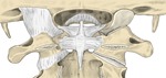
Ligaments of the craniovertebral junction (posterior) 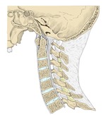
Ligaments of the cervical spine (median section)
Muscle attachments
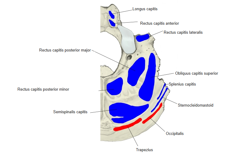
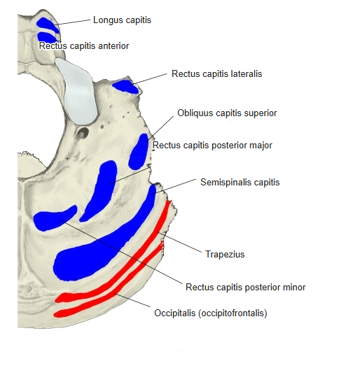
- Origin of: Occipitalis, Trapezius
- Insertion of: Sternocleidomastoid, Splenius capitis, Semispinalis capitis, Rectus capitis posterior major, Rectus capitis posterior minor, Obliquus capitis superior, Rectus capitis lateralis, Rectus capitis anterior, Longus capitis
Trapezius arises from the superior nuchal line and external occipital protuberance; the suboccipital muscles insert below the inferior nuchal line; rectus capitis anterior and longus capitis insert on the basilar part.
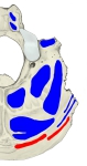
Muscle attachments (from below, version 1) 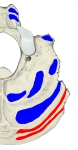
Muscle attachments (from below, version 2) 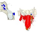
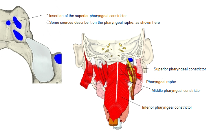
Pharyngeal constrictors
Foramina, fissure and canals
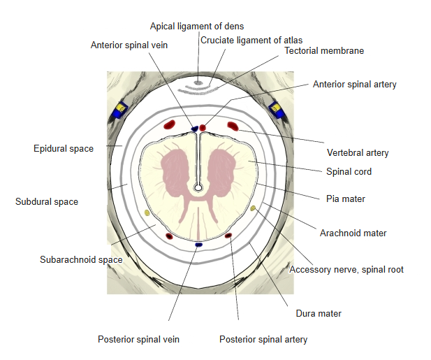
- Foramen magnum – medulla oblongata, spinal roots of the accessory nerve, vertebral arteries, anterior and posterior spinal arteries and veins; the cruciate ligament, apical ligament of dens, tectorial membrane and meninges are related to it.
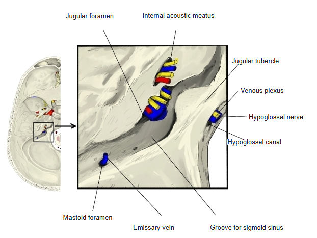
- Jugular foramen (with the temporal bone) – glossopharyngeal, vagus and accessory nerves; internal jugular vein, inferior petrosal sinus; posterior meningeal artery.
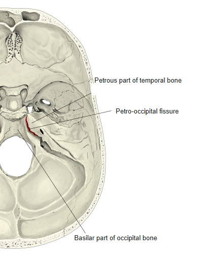
- Petro-occipital fissure – between the posterior border of the petrous part of the temporal bone and the basilar part of the occipital bone.
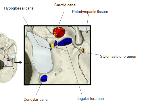
- Condylar canal – opens into the condylar fossa just behind the occipital condyle; transmits the condylar emissary vein.
- Hypoglossal canal – passes through the base of the occipital condyle from back-medial to front-lateral; transmits the hypoglossal nerve and a venous plexus.
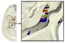
Jugular foramen and hypoglossal canal 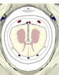
Foramen magnum 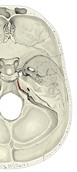
Petro-occipital fissure 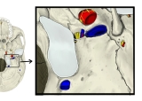
Canals of the cranial base (external)
Development and variation
- The occipital bone forms the back of the cartilaginous skull of the fetus. It arises from four cartilaginous pieces around the foramen magnum and one membrane bone (the upper squama, between the parietal bones).
- Ossification centres appear in the 3rd fetal month; the membranous upper squama fuses early, the other parts later. At about 20 years the occipital bone also fuses with the sphenoid (together called the basi-cranial bone).
- The upper squama may remain separate as an interparietal (Inca) bone, sometimes in two or three pieces.
- Lateral to the condyle the jugular process may project strongly downwards (paramastoid process).
- A small facet on the front edge of the foramen magnum that articulates with the tip of the dens is called a third occipital condyle.
References and sources
- Further reading: Occipital bone, Wikipedia.