Latin: incus (anvil)
Overview
- The middle of the three auditory ossicles; its name means anvil.
- Its body articulates with the head of the malleus in the epitympanic recess, and its long crus carries the lenticular process, which articulates with the head of the stapes. It passes vibrations from the malleus to the stapes.
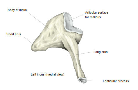
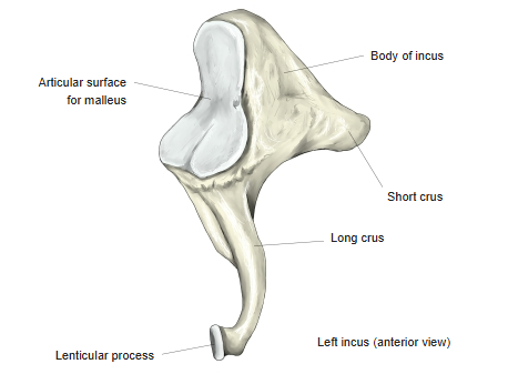
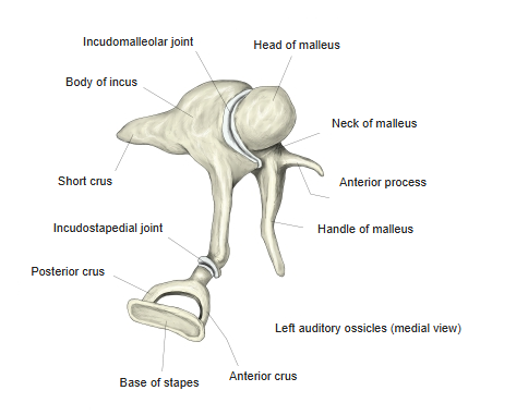
- Its parts are the body, the short crus and the long crus (ending in the lenticular process).
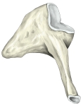
Left incus (medial view) 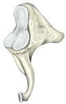
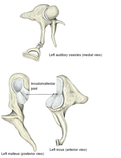
Left incus (anterior view)
- Bones of the skull
- Neurocranium (cranial bones)
- Frontal bone
- Parietal bone (2)
- Occipital bone
- Temporal bone (2)
- Sphenoid bone
- Ethmoid bone
- Viscerocranium (facial bones)
- Nasal bone (2)
- Vomer
- Lacrimal bone (2)
- Inferior nasal concha (2)
- Maxilla (2)
- Zygomatic bone (2)
- Palatine bone (2)
- Mandible
- Hyoid bone
- Neurocranium (cranial bones)
Illustrations
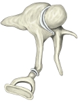
Auditory ossicles (medial view) 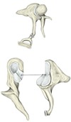
Incudomalleolar joint
Articulations
| # | Bone | Joint |
|---|---|---|
| 1 | Malleus | Incudomalleolar joint (body of incus – head of malleus) |
| 2 | Stapes | Incudostapedial joint (lenticular process – head of stapes) |
Ligaments
The following ligaments attach to the incus. The list is intended to be comprehensive but may omit minor ligaments.
| # | Ligament | Connects |
|---|---|---|
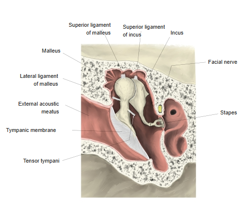
| 1 | Superior ligament of incus | Roof of the epitympanic recess – body |
| 2 | Posterior ligament of incus | Fossa for the incus – short crus |
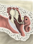
Ligaments of the ossicles 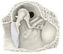
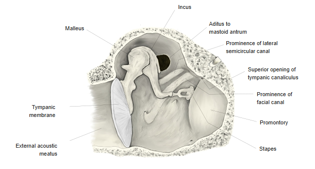
Tympanic cavity (medial view)
Muscle attachments
- Origin of: none
- Insertion of: none
No muscles attach to the incus.
Parts
- Body – lies in the epitympanic recess; its front carries the saddle-shaped articular surface for the malleus.
- Short crus – projects backwards and is anchored by the posterior ligament of the incus in the fossa for the incus, near the aditus to the mastoid antrum.
- Long crus – descends parallel to the handle of the malleus, behind it.
- Lenticular process – a small rounded knob at the tip of the long crus, articulating with the head of the stapes.
References and sources
- Further reading: Incus, Wikipedia.