Middle ear (Latin: auris media)
· Most texts place the tympanic membrane in the external ear, as the wall between the external and middle ear; some sources (such as Wikipedia) count it with the middle ear.
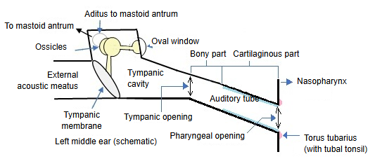
Tympanic cavity: the narrow, air-filled space behind the eardrum. Its medial wall faces the inner ear and has the oval window and the round window. Behind, the aditus to the mastoid antrum opens from its upper part into the mastoid antrum and the mastoid air cells.
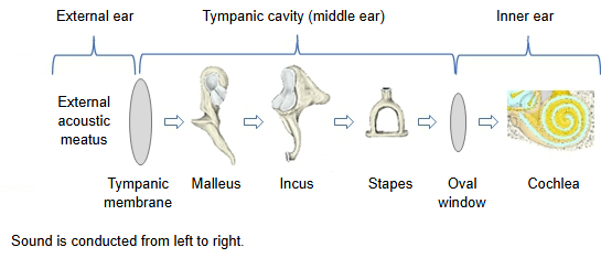
Auditory ossicles: the malleus, incus and stapes, a jointed chain across the cavity from the eardrum to the oval window.
Auditory tube (pharyngotympanic or Eustachian tube): runs forwards, medially and downwards from the cavity to the nasopharynx. Its part near the cavity is bony; the longer part near the pharynx is cartilaginous.
cochlear
organ
- Auditory organ
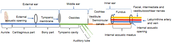
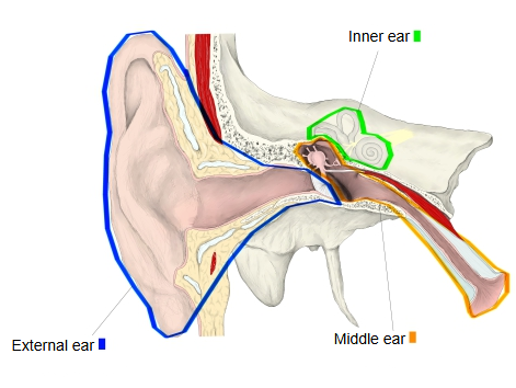
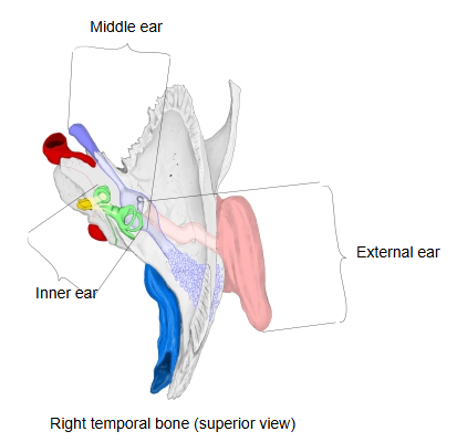
- External ear : 1. auricle, 2. external acoustic meatus, 3. tympanic membrane
- Middle ear : 1. tympanic cavity, 2. auditory ossicles, 3. auditory tube
- Inner ear : its hearing part, the cochlea; the round (cochlear) window closes the basal end of its scala tympani; the internal acoustic meatus is the canal in the temporal bone that carries its nerves and vessels
- Vestibular organ
- Vestibule : part of the inner ear; the oval (vestibular) window opens into it
- Semicircular canals : part of the inner ear: 1. anterior, 2. posterior and 3. lateral semicircular canals
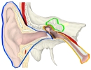 Parts of the ear
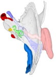 Right temporal bone (superior view)
 External ear to inner ear
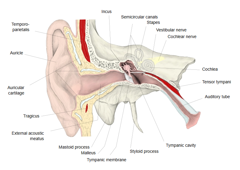
Sound moves the eardrum; the ossicles pass the movement to the base of the stapes, which rocks in the oval window and sets the inner-ear fluid in motion. The eardrum is much larger than the base of the stapes, and the ossicles act as a small lever, so the pressure at the oval window is many times higher than at the eardrum; the vibrations thus reach the fluid without losing most of their energy.
The auditory tube opens during swallowing and yawning and keeps the air pressure in the cavity equal to the pressure outside. Two small muscles damp the chain during loud sounds: the tensor tympani on the malleus and the stapedius on the stapes.
Middle-ear infection (otitis media) is common in young children, partly because their auditory tube is short and nearly horizontal.
Reference websites
[Wikipedia]
· Middle ear, Wikipedia.
· Eustachian tube, Wikipedia.
[Websites with illustrations]
· The middle ear (Kenhub)
· Middle ear (Radiopaedia)
Where it is found
Related terms
- Auditory ossicles
- Tympanic membrane
- Oval window
- Incudostapedial joint
- External acoustic meatus
- Stapes
- Temporal bone
References and sources
- Further reading: Middle ear, Wikipedia.
- Further reading: Eustachian tube, Wikipedia.
- Federative International Programme for Anatomical Terminology (FIPAT). Terminologia Anatomica, 2nd ed. (TA2), 2019.