Latin: malleus (hammer)
Overview
- The outermost and largest of the three auditory ossicles of the middle ear; its name means hammer.
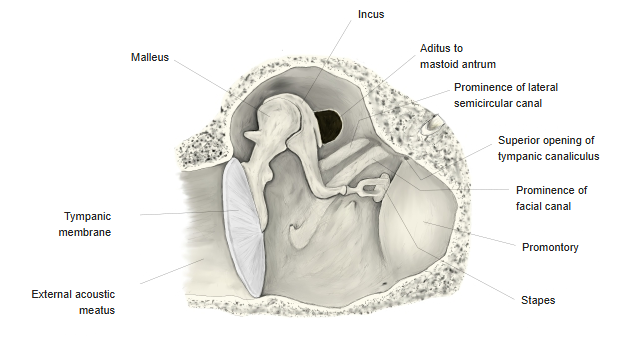
- Its handle is attached to the inner surface of the tympanic membrane, and its head articulates with the incus in the epitympanic recess. Vibrations of the eardrum pass through the malleus to the incus and stapes, and on to the inner ear.
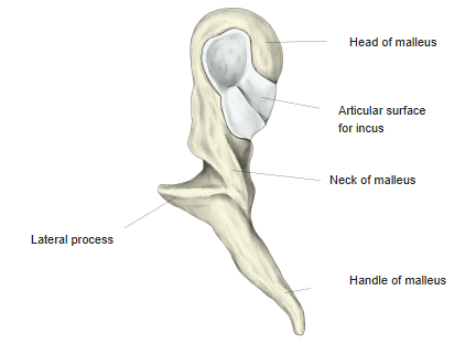
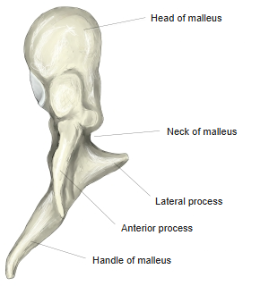
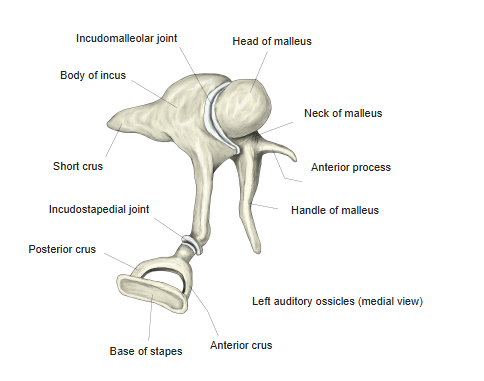
- Its parts are the head, neck, lateral process, anterior process and handle (manubrium).
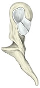
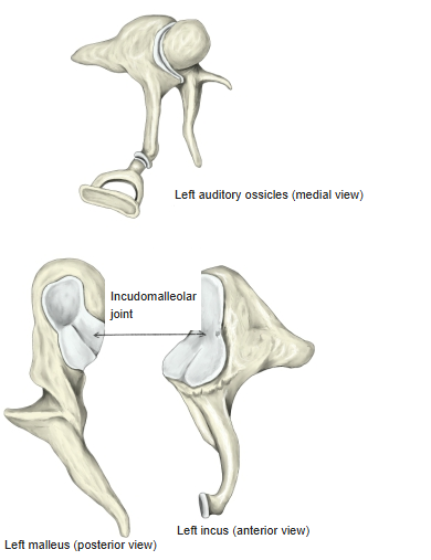
Left malleus (posterior view) 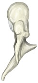
Left malleus (anterior view)
- Bones of the skull
- Neurocranium (cranial bones)
- Frontal bone
- Parietal bone (2)
- Occipital bone
- Temporal bone (2)
- Sphenoid bone
- Ethmoid bone
- Viscerocranium (facial bones)
- Nasal bone (2)
- Vomer
- Lacrimal bone (2)
- Inferior nasal concha (2)
- Maxilla (2)
- Zygomatic bone (2)
- Palatine bone (2)
- Mandible
- Hyoid bone
- Neurocranium (cranial bones)
Illustrations
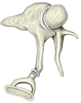
Auditory ossicles (medial view) 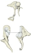
Incudomalleolar joint
Articulations
| # | Bone | Joint |
|---|---|---|
| 1 | Incus | Incudomalleolar joint (saddle-shaped synovial joint) between the head of the malleus and the body of the incus |
Ligaments
The following ligaments attach to the malleus. The list is intended to be comprehensive but may omit minor ligaments.
| # | Ligament | Connects |
|---|---|---|
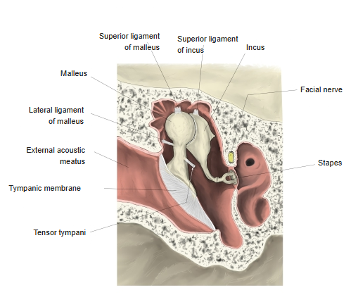
| 1 | Superior ligament of malleus | Roof of the epitympanic recess – head |
| 2 | Anterior ligament of malleus | Anterior process – near the petrotympanic fissure |
| 3 | Lateral ligament of malleus | Edge of the tympanic notch – neck |
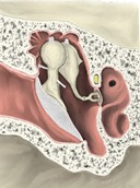
Ligaments of the ossicles 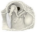
Tympanic cavity (medial view)
Muscle attachments
- Origin of: none
- Insertion of: Tensor tympani
Tensor tympani inserts on the upper part of the handle; by pulling the handle inwards it tenses the tympanic membrane and damps loud sounds.
Parts
- Head – the rounded upper part, lying in the epitympanic recess; its back carries the articular surface for the incus.
- Neck – the narrow part below the head.
- Anterior process – a slender spur running forwards from the neck towards the petrotympanic fissure, held by the anterior ligament.
- Lateral process – a small conical projection pressing on the upper part of the tympanic membrane, where it raises the malleolar prominence.
- Handle (manubrium) – runs downwards and backwards, embedded in the tympanic membrane; its tip reaches the umbo. The tendon of tensor tympani inserts near its root.
References and sources
- Further reading: Malleus, Wikipedia.