Lateral surface of radius (Latin: facies lateralis radii)
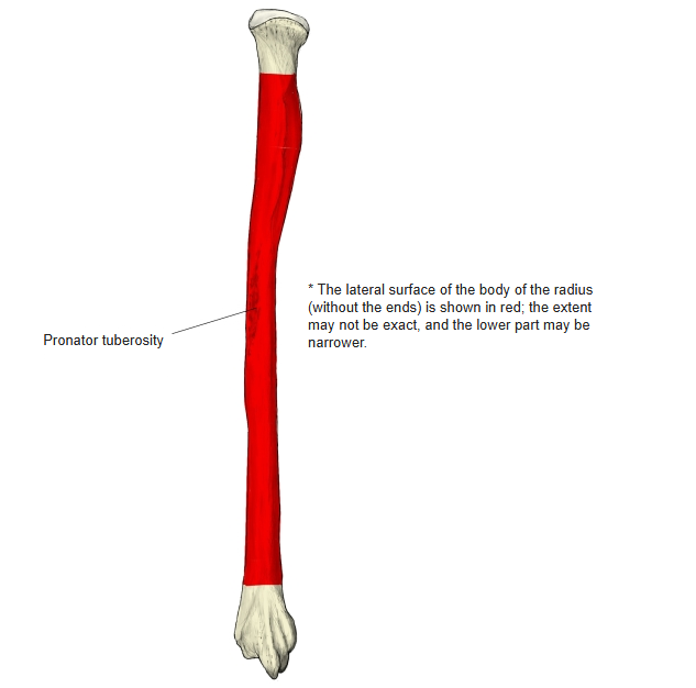
One of the three surfaces of the body (shaft) of the radius: the one that faces laterally (towards the thumb side).
Body of the radius: the shaft between the upper and lower ends of the radius
· It lies between the anterior border and the posterior border; the other two surfaces are the anterior and the posterior surface.
· It is convex (rounded) along its whole length.
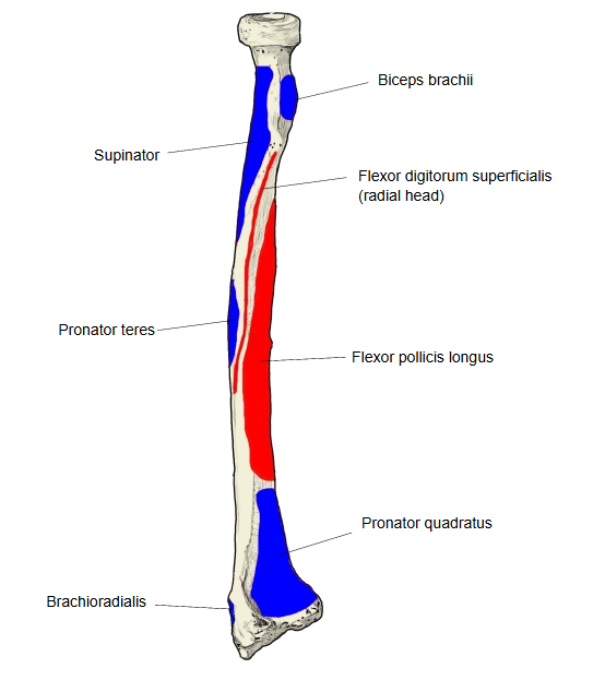
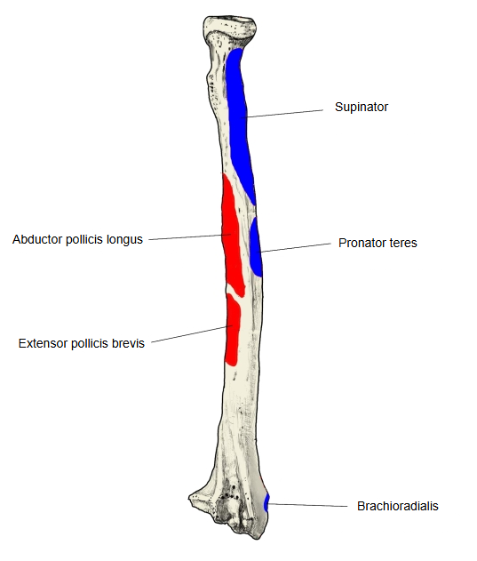
· Its upper third receives the insertion of supinator.
· About its middle, at the point of greatest lateral curvature, is a rough area, the pronator tuberosity, for the insertion of pronator teres.
· Its lower part is narrow and is crossed by the tendons of abductor pollicis longus and extensor pollicis brevis; it ends below at the styloid process, where brachioradialis is inserted.
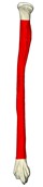 Right radius (lateral)
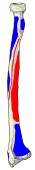 Muscle attachments (anterior)
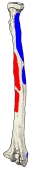 Muscle attachments (posterior)
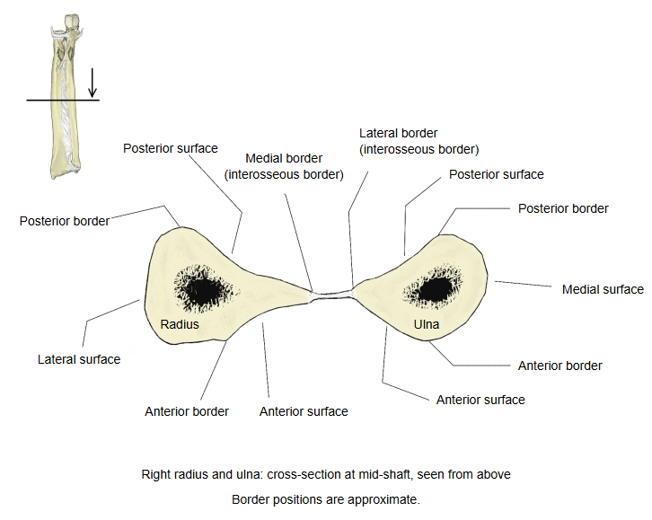 Cross-section of radius and ulna
· Video: The radius - skeleton of the upper limb (the borders and surfaces of the shaft are shown in the video).
Where it is found
Related terms
- Anterior surface of radius
- Posterior surface of radius
- Anterior border of radius
- Posterior border of radius
- Pronator tuberosity
- Styloid process of radius
- Body of radius
References and sources
- Further reading: Radius (bone), Wikipedia.
- Further reading: Supinator muscle, Wikipedia.
- Further reading: Pronator teres muscle, Wikipedia.
- Federative International Programme for Anatomical Terminology (FIPAT). Terminologia Anatomica, 2nd ed. (TA2), 2019.