Trochanteric fossa (Latin: fossa trochanterica)
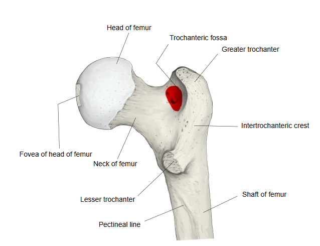
A deep pit seen when the upper end of the femur is viewed from behind. It lies on the medial side of the greater trochanter, between the overhanging top of the trochanter and the root of the neck of the femur. The tendon of obturator externus is inserted into it; obturator internus and the superior and inferior gemelli are inserted just above it.
Sources differ: some place the insertions of obturator internus and the gemelli in the fossa itself. Fibres of the ischiofemoral ligament pass behind the neck and end close to the fossa. Surgeons sometimes call this area the piriformis fossa, although piriformis is inserted on the upper border of the greater trochanter; it is a landmark for inserting nails into the femoral shaft.
Attachments
| Muscle | 1 | Obturator internus (insertion, just above the fossa) | musculus obturatorius internus |
| 2 | Obturator externus (insertion) | musculus obturatorius externus | |
| 3 | Gemellus superior (insertion, with obturator internus) | musculus gemellus superior | |
| 4 | Gemellus inferior (insertion, with obturator internus) | musculus gemellus inferior | |
| Ligament | 1 | Ischiofemoral ligament (fibres end close to the fossa) | ligamentum ischiofemorale |
Reference websites
[Wikipedia]
· Trochanteric fossa, Wikipedia.
· External obturator muscle, Wikipedia.
[Websites with illustrations]
· Upper end of the femur, posterior view (Wikimedia Commons)

· Right femur, posterior surface (Gray's Anatomy, fig. 245) (Wikimedia Commons)

· The femur (TeachMeAnatomy)
Where it is found
Related terms
- Greater trochanter
- Neck of femur
- Intertrochanteric crest
- Lesser trochanter
- Proximal epiphysis of femur
- Hip joint
References and sources
- Further reading: Trochanteric fossa, Wikipedia.
- Further reading: External obturator muscle, Wikipedia.
- Federative International Programme for Anatomical Terminology (FIPAT). Terminologia Anatomica, 2nd ed. (TA2), 2019.