Lesser trochanter (Latin: trochanter minor)
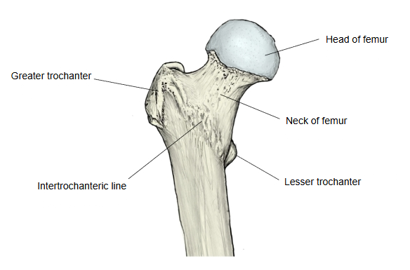
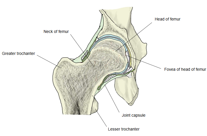
A small conical projection of the femur, best seen when the upper end is viewed from behind. It lies at the junction of the neck of femur and the shaft of femur and points medially and backwards. Behind, it is joined to the greater trochanter by the intertrochanteric crest; in front, the intertrochanteric line runs towards it but passes below it as the spiral line, and the pectineal line runs down from its base to the linea aspera. Its rough tip receives the tendon of iliopsoas (psoas major and iliacus), the strongest flexor of the hip joint, and the pubofemoral ligament blends with the joint capsule just above it (sources differ on whether its fibres reach the trochanter itself). Avulsion of the lesser trochanter by iliopsoas is seen in adolescent athletes; in adults an isolated avulsion is often a sign of a tumour in the bone.
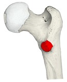 Right femur, upper end (posterior)
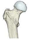 Right femur, upper end (anterior)
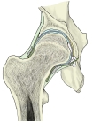 Right hip joint (coronal section)
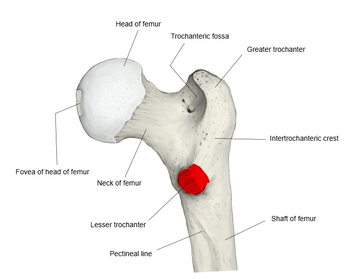
Attachments
| Muscles | 1 | psoas major | m. psoas major |
| 2 | iliacus | m. iliacus | |
| Ligament | 1 | pubofemoral ligament | lig. pubofemorale |
Where it is found
Related terms
- Greater trochanter
- Intertrochanteric crest
- Intertrochanteric line
- Neck of femur
- Pectineal line of femur
- Proximal epiphysis of femur
- Hip joint
References and sources
- Further reading: Lesser trochanter, Wikipedia.
- Further reading: Iliopsoas, Wikipedia.
- Federative International Programme for Anatomical Terminology (FIPAT). Terminologia Anatomica, 2nd ed. (TA2), 2019.