Posterior surface of tibia (Latin: facies posterior tibiae)
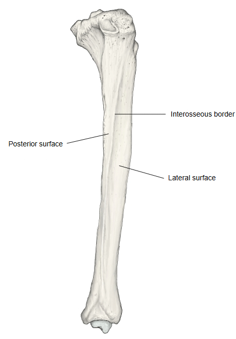
Of the three surfaces of the shaft of the tibia, the one that faces backwards, between the medial border and the interosseous border. Its upper part (about the upper third) is crossed by the soleal line.
tibia
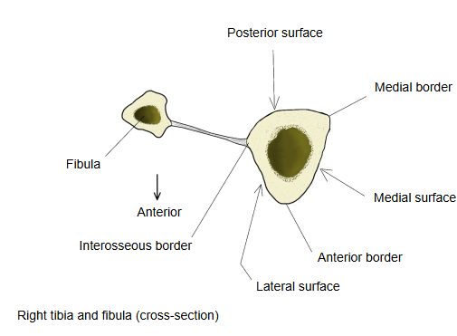
- Medial surface : the surface between the medial border and the anterior border.
- Lateral surface : the surface between the anterior border and the interosseous border.
- Posterior surface : the surface that faces backwards.
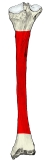 Right tibia (posterior)
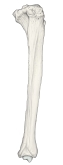 Right tibia (lateral)
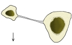 Tibia and fibula in cross-section
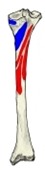 Muscle attachments (posterior)
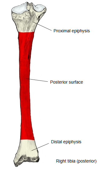
It is widest above. The soleal line runs obliquely down and medially across its upper part, from the back of the fibular articular facet to the medial border at the junction of the upper and middle thirds. The triangular area above the line receives the insertion of popliteus; the line itself gives origin to part of soleus.
Below the line, a faint vertical line divides the middle third into two parts: flexor digitorum longus arises from the broader medial part and tibialis posterior from the narrower lateral part. A large nutrient foramen, directed downwards, opens just below the soleal line. The lower third is smooth and is covered by tibialis posterior, flexor digitorum longus and flexor hallucis longus.
Attachments
| Muscles | 1 | Flexor digitorum longus (origin) | medial part of the middle third, below the soleal line | 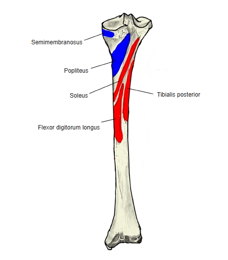 |
| 2 | Soleus (origin) | soleal line and the medial border below it | ||
| 3 | Tibialis posterior (origin) | lateral part of the middle third, below the soleal line | ||
| 4 | Popliteus (insertion) | triangular area above the soleal line |
Reference websites
[Wikipedia]
· Tibia, Wikipedia.
· Soleus muscle, Wikipedia.
[Websites with illustrations]
· Bones of the right leg, posterior surface (Gray's Anatomy, fig. 259) (Wikimedia Commons)

· Tibia: anatomy and clinical notes (Kenhub)
· The tibia: proximal, shaft and distal anatomy (TeachMeAnatomy)
Where it is found
Related terms
- Soleal line
- Medial border of tibia
- Interosseous border of tibia
- Lateral surface of tibia
- Medial surface of tibia
- Shaft of tibia
References and sources
- Further reading: Tibia, Wikipedia.
- Further reading: Soleus muscle, Wikipedia.
- Federative International Programme for Anatomical Terminology (FIPAT). Terminologia Anatomica, 2nd ed. (TA2), 2019.