Shaft of tibia (Latin: corpus tibiae)
Also called Body of tibia
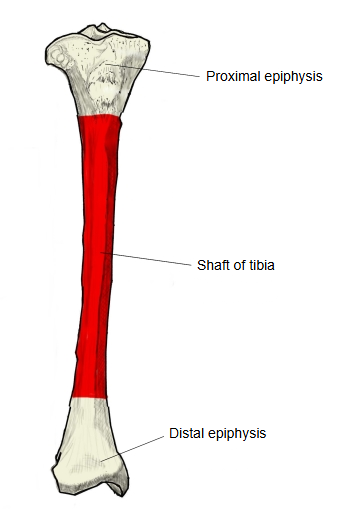
The shaft (body) of the tibia is the long middle part of the tibia, the diaphysis that remains when the bone is divided into three parts and the broad upper and lower ends are left out.
It is roughly a three-sided prism. It is thinnest at the junction of its middle and lower thirds, a common site of tibial fractures. Its lower part turns a little laterally, towards the fibula, so that seen from the front the shaft has a slight S-shaped curve. It has three borders (anterior, medial and interosseous) and three surfaces (medial, lateral and posterior).
Its anterior border and medial surface lie just under the skin and form the shin. Because the shin has so little soft-tissue cover, fractures of the tibial shaft are often open fractures (tibial fracture).
- Proximal epiphysis : much larger than the shaft; the upper end.
- Shaft of tibia : the diaphysis (body) of the bone.
- Distal epiphysis : expanded, but less than the upper end; the lower end.
Three surfaces and three borders can be distinguished.
- Anterior border : the sharpest of the three, especially above; it runs from the tibial tuberosity to the front of the medial malleolus with a gentle S-shaped curve.
- Medial border : rounded; it runs from the medial condyle to the back of the medial malleolus.
- Interosseous border : a thin ridge facing the fibula that splits into two lines below (bounding the fibular notch); the interosseous membrane of the leg is attached to it.
- Medial surface : a broad, smooth surface not covered by muscle; it can be felt, together with the anterior border, along its whole length.
- Lateral surface : narrower than the medial surface and smooth; its upper part is a shallow hollow from which tibialis anterior arises.
- Posterior surface : crossed above by the oblique soleal line; a large nutrient foramen lies just below and lateral to it, and a faint vertical line runs down from there.
Besides the borders and surfaces, the following features are found:
| 1 | Tibial tuberosity | tuberositas tibiae | |
| 2 | Soleal line | linea musculi solei |
Attached structures
| Muscles | 1 | Tibialis posterior (origin) | 8 | Rectus femoris (insertion, via the patellar ligament) | Right tibia Anterior Posterior 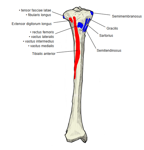 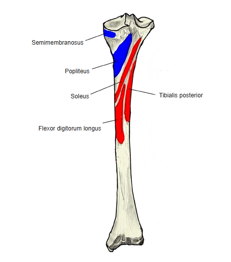 |
| 2 | Tibialis anterior (origin) | 9 | Vastus intermedius (insertion, via the patellar ligament) | ||
| 3 | Flexor digitorum longus (origin) | 10 | Vastus medialis (insertion, via the patellar ligament) | ||
| 4 | Extensor digitorum longus (origin, from the lateral condyle above the shaft) | 11 | Gracilis (insertion) | ||
| 5 | Soleus (origin) | 12 | Semitendinosus (insertion) | ||
| 6 | Vastus lateralis (insertion, via the patellar ligament) | 13 | Sartorius (insertion) | ||
| 7 | Popliteus (insertion) | ||||
| Ligaments | 1 | Patellar ligament | 2 | Tibial collateral ligament |
Origins: tibialis anterior from the lateral surface; tibialis posterior and flexor digitorum longus from the posterior surface, on either side of the vertical line; soleus from the soleal line. Insertions: popliteus on the posterior surface above the soleal line; sartorius, gracilis and semitendinosus on the upper medial surface (pes anserinus). The patellar ligament attaches to the tibial tuberosity at the top of the anterior border and carries the pull of quadriceps femoris; the tibial collateral ligament attaches to the upper medial surface.
Where it is found
Related terms
- Anterior border of tibia
- Medial border of tibia
- Interosseous border of tibia
- Medial surface of tibia
- Lateral surface of tibia
- Posterior surface of tibia
- Soleal line
References and sources
- Further reading: Tibia, Wikipedia.
- Further reading: Tibial fracture, Wikipedia.
- Federative International Programme for Anatomical Terminology (FIPAT). Terminologia Anatomica, 2nd ed. (TA2), 2019.