Tibiofibular syndesmosis (Latin: syndesmosis tibiofibularis)
Also called the inferior (distal) tibiofibular joint
The fibular notch, on the lateral side of the lower end of the tibia, and the medial side of the lower end of the fibula meet with rough surfaces and no joint cavity between them. The union is strengthened in front by the anterior tibiofibular ligament and behind by the posterior tibiofibular ligament. This kind of union is called the tibiofibular syndesmosis.
· It is a syndesmosis, a fibrous joint in which the bones are held together by strong ligaments. It is also called the inferior (distal) tibiofibular joint; the upper joint between the two bones is the tibiofibular joint.
· It holds the lateral malleolus firmly against the tibia, keeping the socket of the ankle (the ankle mortise) tight around the trochlea of the talus. It allows only slight movement: the fibula moves a little as the wider front of the talus enters the mortise when the foot is pulled up (dorsiflexion).
There is no joint cavity here, but a small pouch of the cavity of the talocrural (ankle) joint may extend a short way up between the two bones.
 Right ankle (section)
 Right ankle (lateral)
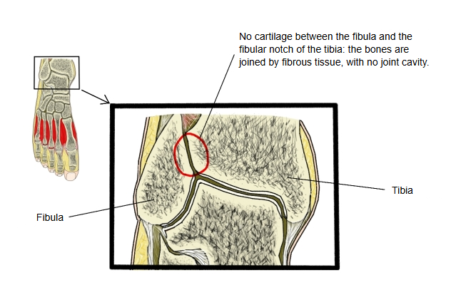
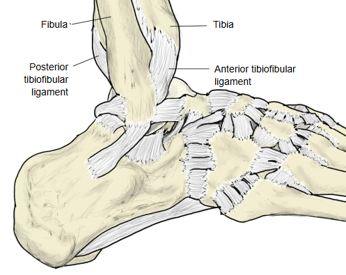
Some sources also count the interosseous membrane of the leg among the ligaments that strengthen this joint.
The joint surfaces are the rough, concave fibular notch of the tibia and the rough, convex medial side of the lower end of the fibula, just above the articular surface of the lateral malleolus. The lowest few millimetres (about 4 mm) of these surfaces may be smooth and covered with cartilage continuous with that of the ankle joint; sources differ on this. Between the rough surfaces runs the interosseous ligament, short strong fibres continuous above with the interosseous membrane. The deep part of the posterior tibiofibular ligament is the transverse tibiofibular ligament, which deepens the back of the socket for the talus.
Injury to these ligaments is called a high ankle sprain; the syndesmosis is also often damaged in ankle fractures.
Reference websites
[Wikipedia]
· Inferior tibiofibular joint, Wikipedia.
· Syndesmosis, Wikipedia.
· Ankle, Wikipedia.
[Websites with illustrations]
· Coronal section through the right ankle (Gray's Anatomy) (Wikimedia Commons)

· Ligaments of the foot, lateral aspect (Gray's Anatomy) (Wikimedia Commons)

· Tibiofibular joints (Kenhub)
Where it is found
Related terms
- Fibular notch
- Talocrural joint
- Lateral malleolus
- Distal epiphysis of fibula
- Distal epiphysis of tibia
- Tibiofibular joint
References and sources
- Further reading: Syndesmosis, Wikipedia.
- Further reading: Ankle, Wikipedia.
- Federative International Programme for Anatomical Terminology (FIPAT). Terminologia Anatomica, 2nd ed. (TA2), 2019.