Distal epiphysis of tibia (Latin: epiphysis distalis tibiae)
Also called the lower end of tibia
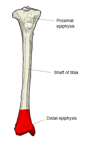
The distal epiphysis is the expanded lower part of the tibia when the bone is divided into three parts; it is also called the lower end. It forms the roof and inner wall of the ankle joint.
It is expanded, but less so than the upper end. On its medial side it extends downwards as the medial malleolus; the smooth lateral face of the malleolus is the articular surface of the medial malleolus, part of the joint surface for the talus. The lower surface is the smooth inferior articular surface for the talus, concave from front to back and slightly convex from side to side, divided in two by a low ridge running from front to back.
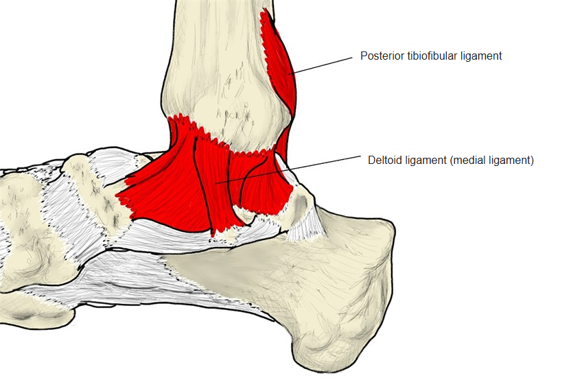
Behind the medial malleolus is the malleolar groove for the tendons of tibialis posterior and flexor digitorum longus. On the lateral side is the fibular notch, a hollow that holds the lower end of the fibula at the tibiofibular syndesmosis; the anterior, posterior and transverse tibiofibular ligaments attach around it, and the deltoid ligament arises from the medial malleolus. Strictly, an epiphysis is the part beyond the growth plate in a growing bone; here the term is used for the whole lower end of the adult tibia.
- Proximal epiphysis : much larger than the shaft; the upper end.
- Shaft of tibia : the diaphysis (body) of the bone.
- Distal epiphysis : expanded, but less than the upper end; the lower end.
The following features are found:
| 1 | Medial malleolus | malleolus medialis | 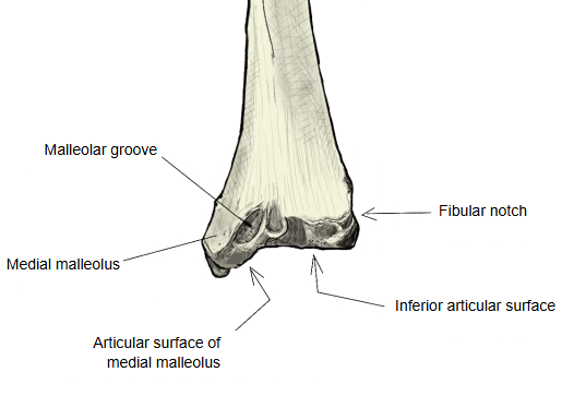 |
| 2 | Articular surface of medial malleolus | facies articularis malleoli medialis | |
| 3 | Malleolar groove | sulcus malleolaris | |
| 4 | Fibular notch | incisura fibularis | |
| 5 | Inferior articular surface | facies articularis inferior |
Attached structures
| Ligaments | 1 | Anterior tibiofibular ligament | ligamentum tibiofibulare anterius |
| 2 | Posterior tibiofibular ligament | ligamentum tibiofibulare posterius | |
| 3 | Transverse tibiofibular ligament | ligamentum tibiofibulare transversum | |
| 4 | Deltoid ligament | ligamentum deltoideum (ligamentum collaterale mediale) | |
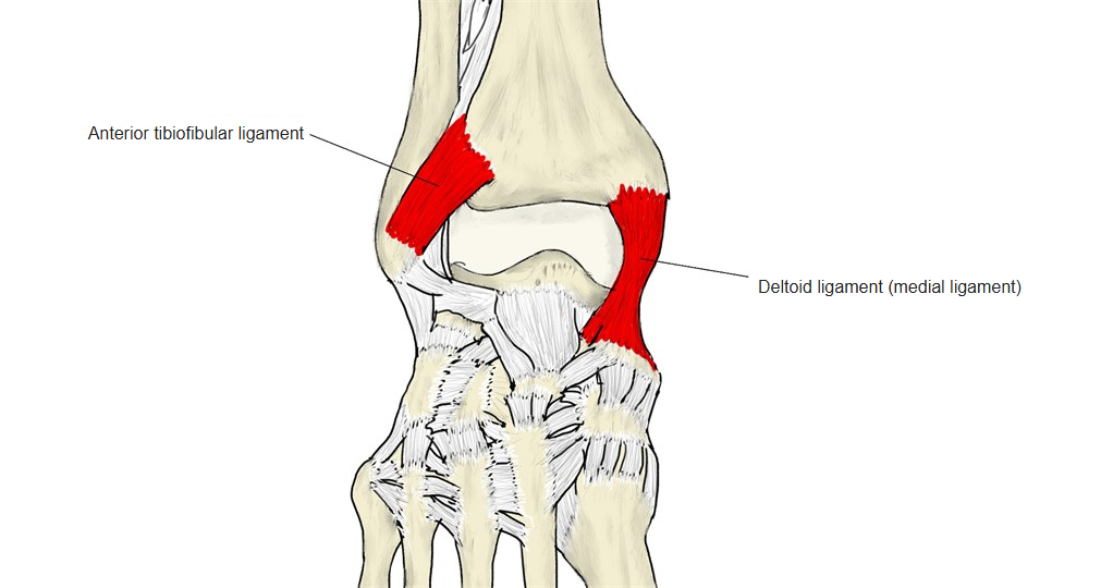 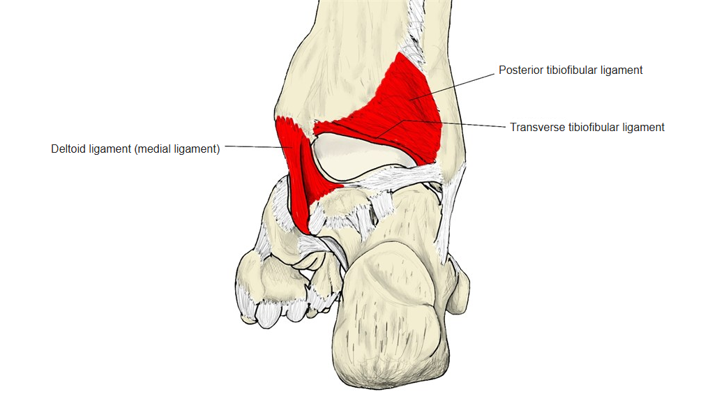  | |||
Reference websites
[Wikipedia]
· Tibia, Wikipedia.
· Ankle, Wikipedia.
[Websites with illustrations]
· Tibia, inferior epiphysis (posterior view) (Wikimedia Commons)

· Tibia, inferior epiphysis (anterior view) (Wikimedia Commons)

· The tibia (TeachMeAnatomy)
Where it is found
Related terms
- Medial malleolus
- Articular surface of medial malleolus
- Malleolar groove of tibia
- Fibular notch
- Inferior articular surface of tibia
- Shaft of tibia
- Talocrural joint