Latin: os capitatum · one of the carpal bones of the hand
Overview
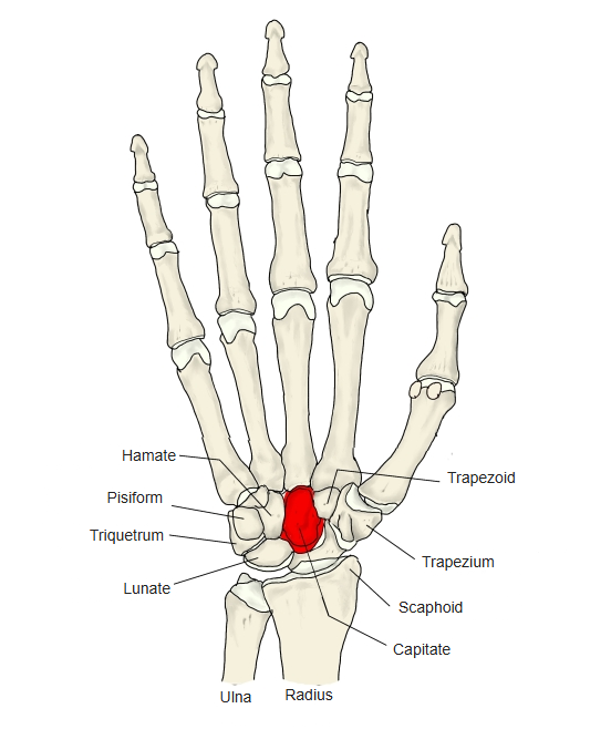
- One of the eight carpal bones; second from the little-finger side in the distal row.
- The largest carpal bone, with four articular surfaces (superior, inferior, medial and lateral).
- Its upper end forms a rounded head that fits against the scaphoid and lunate and lies at the centre of the carpus.
- Its lower end articulates mainly with the third metacarpal, and also with the second and fourth.
- Begins to ossify at about 1–5 months of age.
- Carpal bones (8)
- Distal row (4)
- Trapezium ← thumb side
- Trapezoid
- Capitate
- Hamate ← little-finger side
- Proximal row (4)
- Scaphoid ← thumb side
- Lunate
- Triquetrum
- Pisiform ← little-finger side
- Distal row (4)
Illustrations
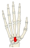
Bones of the hand (palmar view) 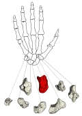
Carpal bones drawn separately (palmar view) 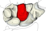
Carpal bones (palmar view) 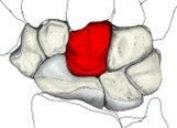
Carpal bones (dorsal view) 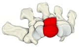
Carpal bones and metacarpals 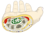
Wrist (cross-section) 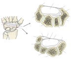
Flexor retinaculum (right)
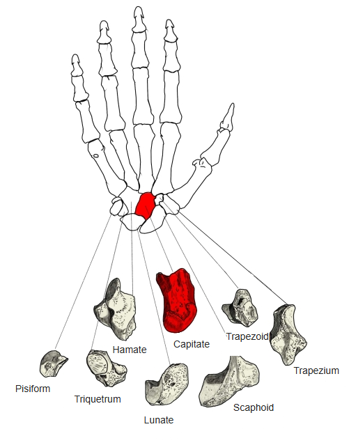
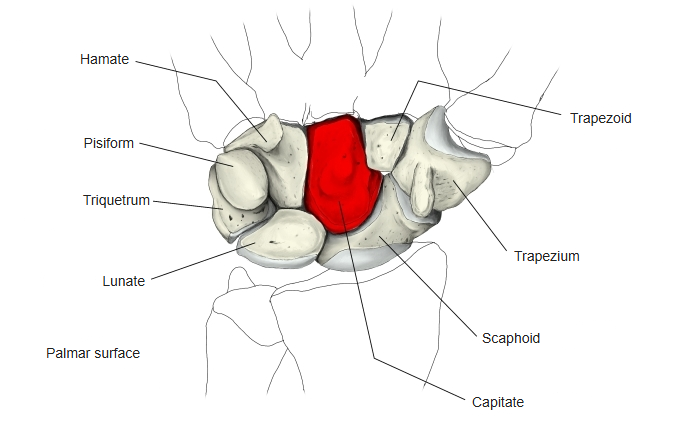
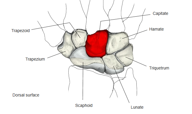
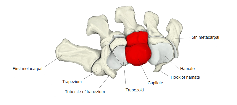
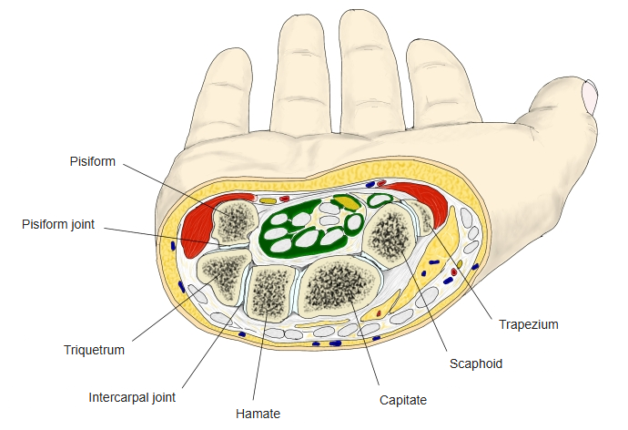
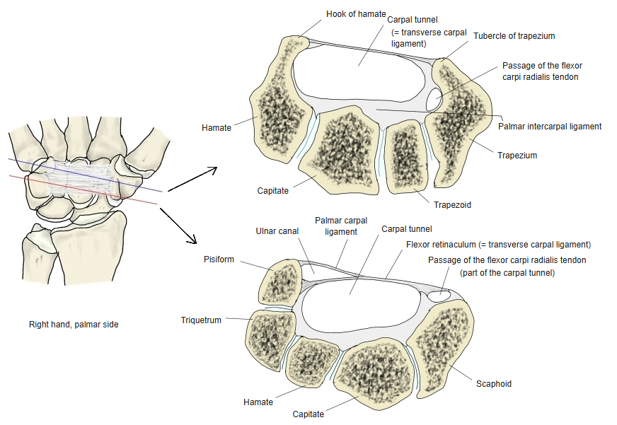
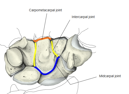
Articulations
Ligaments
The following ligaments attach to the capitate. The list is intended to be comprehensive but may omit minor ligaments.
| # | Ligament | Connects |
|---|---|---|
| 1 | Interosseous intercarpal ligaments | Scaphoid – lunate – triquetrum; trapezium – trapezoid – capitate – hamate |
| 2 | Dorsal intercarpal ligaments | Between the carpal bones |
| 3 | Palmar intercarpal ligaments | Between the carpal bones |
| 4 | Radiate carpal ligament | Capitate → the surrounding carpal bones |
| 5 | Dorsal carpometacarpal ligaments | Distal carpal bones – metacarpals (except the first) |
| 6 | Palmar carpometacarpal ligaments | Distal carpal bones – metacarpals |
Muscle attachments
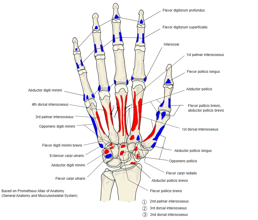
- Origin of: Adductor pollicis, Flexor pollicis brevis
- Insertion of: none
The capitate gives origin to the oblique head of the adductor pollicis and the deep head of the flexor pollicis brevis.
Muscle attachments of the hand (palmar) 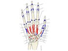
Muscle attachments of the hand
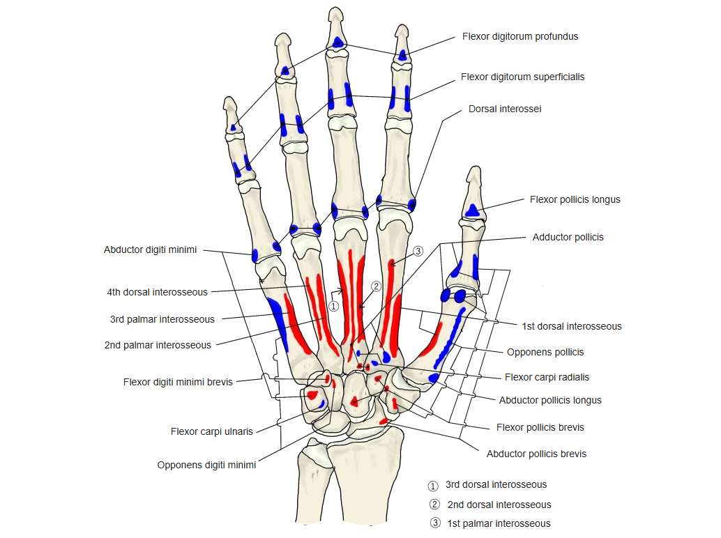
Surfaces
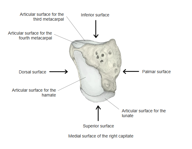
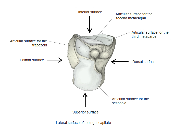
- Superior surface: round and smooth; articulates with the lunate.
- Inferior surface: divided by two ridges into three facets, for the second, third and fourth metacarpals.
- Dorsal surface: broad and rough.
- Palmar surface: narrow, rounded and rough, for ligaments and part of the adductor pollicis.
- Lateral surface: a small facet for the trapezoid, an area for an interosseous intercarpal ligament, and a convex facet for the scaphoid.
- Medial surface: an oblong concave facet for the hamate; rough in front for an interosseous intercarpal ligament.
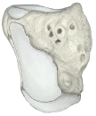
Right hand, medial side 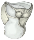
Right hand, lateral side
References and sources
- Further reading: Capitate bone, Wikipedia.