Latin: clavicula · also called the collarbone
Overview
- One of the two bones of the shoulder girdle (with the scapula).
- It lies almost horizontally across the front of the body, at the base of the neck and the front of the shoulder; it can easily be felt through the skin.
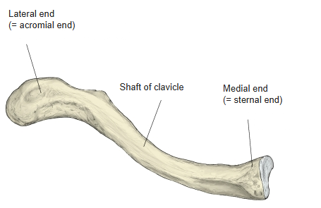
- A rod-shaped long bone with an S-shaped curve.
- It has a shaft and two slightly enlarged ends — the acromial (lateral) end and the sternal (medial) end — each with an articular facet.
- It holds the scapula away from the trunk, linking the upper limb to the axial skeleton.
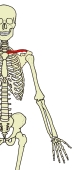
Bones of the upper limb 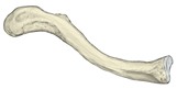
Parts of the clavicle
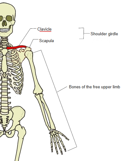
- Bones of the upper limb (one side)
- Shoulder girdle
- Clavicle
- Scapula
- Free upper limb
- Arm
- Humerus
- Forearm
- Radius ← thumb side
- Ulna ← little-finger side
- Hand
- Carpal bones (8)
- Metacarpal bones (5)
- Phalanges (14)
- Arm
- Shoulder girdle
Illustrations
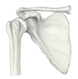
Position of the clavicle (anterior view) 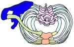
Position of the clavicle (superior view)
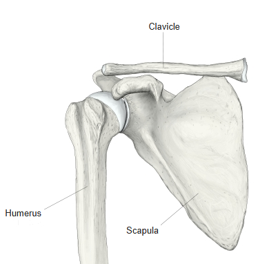
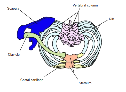
Articulations
| # | Bone | Joint |
|---|---|---|
| 1 | Scapula | Acromioclavicular joint |
| 2 | Sternum | Sternoclavicular joint |
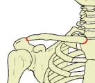
Joints of the clavicle
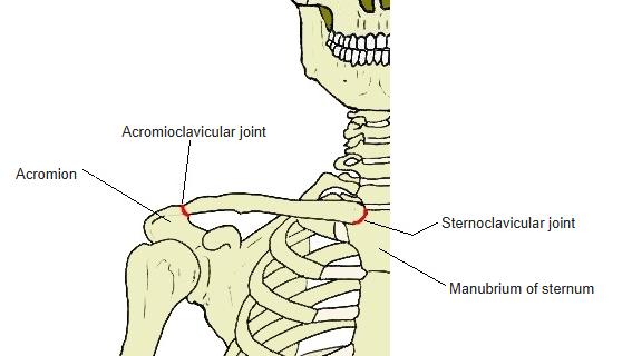
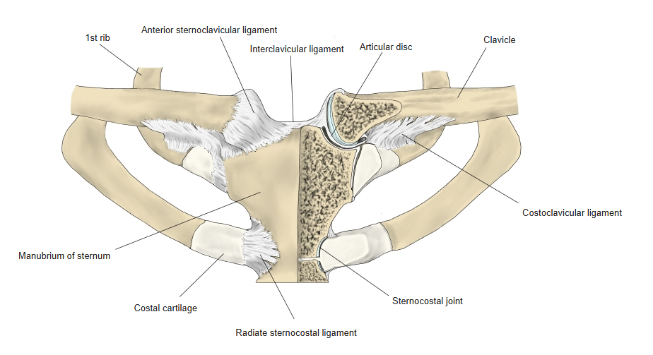
Acromioclavicular joint: acromial facet of the clavicle ↔ clavicular facet of the acromion. Sternoclavicular joint: sternal facet of the clavicle ↔ clavicular notch of the manubrium (with an articular disc); the first costal cartilage also forms part of this joint.
Ligaments
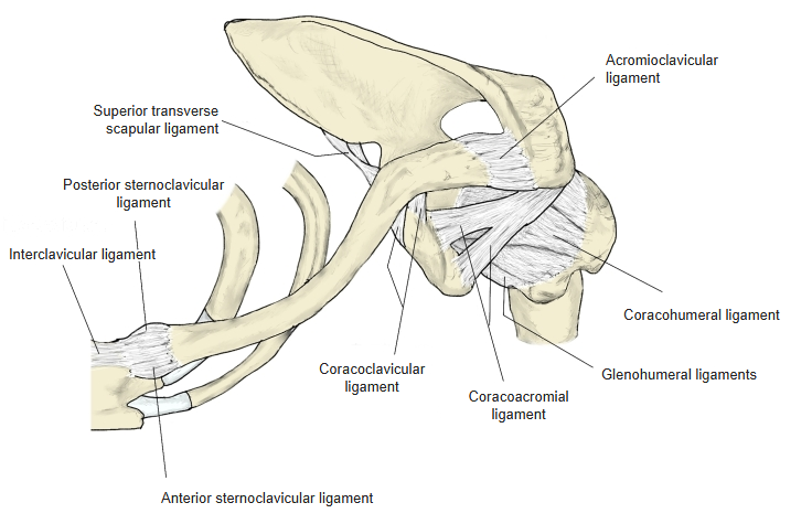
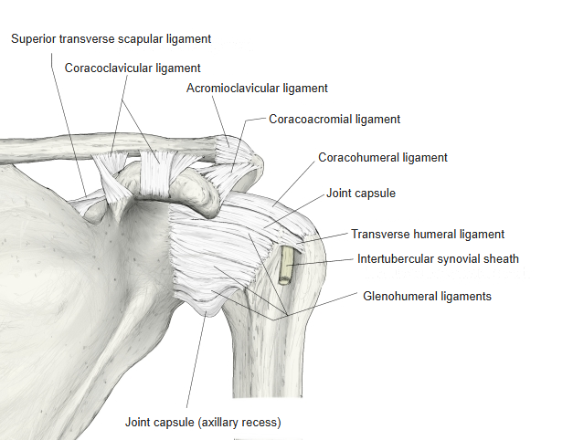
The following ligaments attach to the clavicle. The list is intended to be comprehensive but may omit minor ligaments.
| # | Ligament | Connects |
|---|---|---|
| 1 | Coracoclavicular ligament — conoid ligament and trapezoid ligament | Coracoid process – lateral part of the shaft (conoid tubercle, trapezoid line) |
| 2 | Acromioclavicular ligament | Acromion – acromial end |
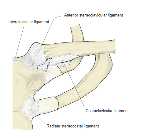
| 3 | Anterior sternoclavicular ligament | Front of the sternal end – front of the manubrium |
| 4 | Posterior sternoclavicular ligament | Back of the sternal end – back of the manubrium |
| 5 | Interclavicular ligament | Between the upper parts of the sternal ends of both clavicles |
| 6 | Costoclavicular ligament | Impression for the costoclavicular ligament – 1st rib |
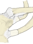
Sternoclavicular joint ligaments 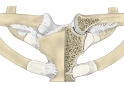
Sternoclavicular joints (section) 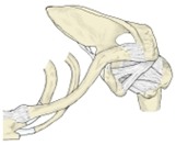
Ligaments around the shoulder region (from above) 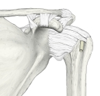
Right shoulder joint (anterior)
Muscle attachments
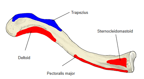
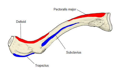
- Origin of: Sternocleidomastoid, Deltoid, Pectoralis major
- Insertion of: Subclavius, Trapezius
Origins: sternocleidomastoid — upper surface of the medial third; deltoid — anterior border and upper surface of the lateral third; pectoralis major — anterior surface of the medial half. Insertions: subclavius — subclavian groove; trapezius — posterior border and upper surface of the lateral third.
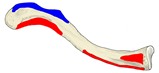
Muscle attachments (upper surface) 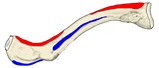
Muscle attachments (lower surface) 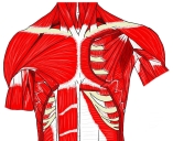
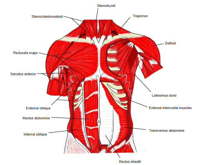
Muscles of the trunk (anterior)
Development and structure
- A membrane (dermal) bone: it is the first bone of the body to start ossifying (5th fetal week), but the last long bone to finish (after about 25 years).
- Unlike other long bones it has no medullary cavity; the inside is filled with spongy bone.
- Well developed in mammals that use the forelimbs for more than walking (grasping, crossing the arms in front of the body), such as primates and rodents; absent or vestigial in those that use them only for walking, such as carnivores and ungulates.
Variations
- Its shape varies more than that of most other long bones.
- It is occasionally pierced by a branch of a supraclavicular nerve.
- In males it is thicker and more curved, with more pronounced muscle attachments; in females it is thinner, smoother and lighter. Its form is a reliable criterion for sex determination.
- The right clavicle is usually stronger and shorter than the left.
- It may be partly or completely absent in cleidocranial dysplasia.
- A levator claviculae muscle (in about 2–3% of people) runs from the transverse processes of the upper cervical vertebrae to the lateral half of the clavicle.
Fractures
- Because it holds the scapula away from the trunk, it breaks easily in a fall onto the shoulder; the force is usually indirect, through the hand or shoulder.
- Most fractures occur about one third of the way from the lateral end, where the two curves meet — the weakest part (muscle attachments may also play a role). The weight of the arm can displace the fragments.
- Fractures of the medial part also occur, but the fragments are not displaced.
Blood supply
- Mainly from the suprascapular artery and branches of the thoracoacromial artery; branches of the internal thoracic artery also reach the sternal end (sternoclavicular joint).
- The suprascapular artery runs laterally along the upper surface and gives a branch that enters the nutrient foramen near the junction of the lateral and middle thirds.
- Two branches of the thoracoacromial artery pierce the attachments of the pectoralis major and deltoid to reach the periosteum of the middle third; they often anastomose between the attachments.
- The blood supply is mainly periosteal; the intraosseous arteries are few and short. Periosteal vessels are found on the upper surface and anterior border, not on the lower surface or posterior border.
- Extensive stripping of the periosteum during surgery for a fracture may therefore lead to non-union.
Etymology
- Latin clavicula, the diminutive of clavis (“key”, “bolt”): a “little key”.
References and sources
- Havet E, et al. Vascular anatomical basis of clavicular non-union. Surgical and Radiologic Anatomy, 2008 — blood supply.
- Further reading: Clavicle, Wikipedia.