Zygomaticotemporal foramen (Latin: foramen zygomaticotemporale)
A small opening on the temporal surface of the zygomatic bone.
bone
- Lateral surface
- Orbital surface
- Temporal surface
- Zygomaticotemporal foramen : the zygomaticotemporal nerve comes out
The back part of the inner side of the zygomatic bone is the temporal surface. It faces backwards and medially and forms the front wall of the temporal fossa; the zygomaticotemporal foramen is on this surface, and the zygomaticotemporal nerve leaves the bone through it.
It is usually described as single and in the middle of the temporal surface. It is one of the exits of the zygomatic canal, which starts at the zygomatico-orbital foramen on the orbital surface; the other exit is the zygomaticofacial foramen on the lateral surface. The zygomaticotemporal nerve is a branch of the zygomatic nerve (from the maxillary nerve) and supplies the skin of the temple.
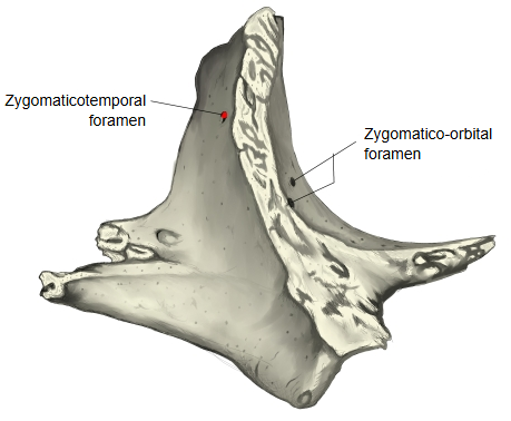
Question
On its position: textbooks and Wikipedia place it in the centre of the temporal surface, but in many published illustrations it does not seem to lie in the centre. There is individual variation, but it seems more often to open higher, above the level of the zygomatico-orbital foramen.
Reference websites
[Wikipedia]
· Zygomaticotemporal foramen, Wikipedia.
· Zygomaticotemporal nerve, Wikipedia.
[Websites with illustrations]
· Left zygomatic bone, inner surface (Gray's Anatomy, fig. 166) (Wikimedia Commons)

· Zygomatic bone (Kenhub)
⇒ Back to the zygomatic bone page
Where it is found
Related terms
References and sources
- Further reading: Zygomaticotemporal foramen, Wikipedia.
- Further reading: Zygomaticotemporal nerve, Wikipedia.
- Federative International Programme for Anatomical Terminology (FIPAT). Terminologia Anatomica, 2nd ed. (TA2), 2019.