Zygomatic arch (Latin: arcus zygomaticus)
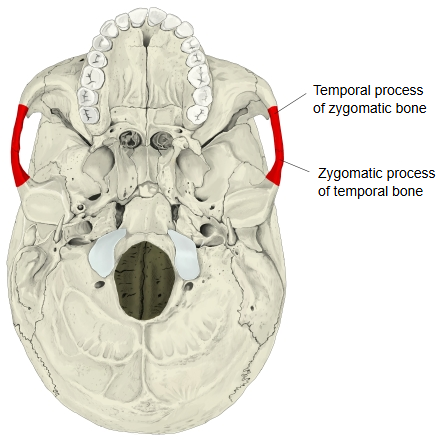
Zygomatic bone (temporal process) + temporal bone (zygomatic process) → zygomatic arch
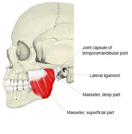
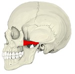 Skull (left lateral view)
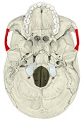 External surface of cranial base
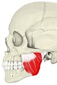 Masseter
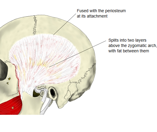
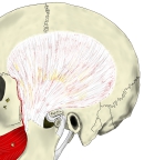 Temporal fascia
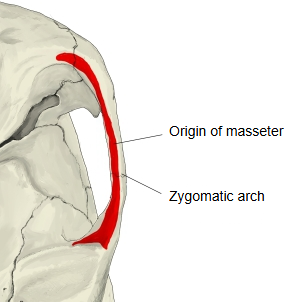
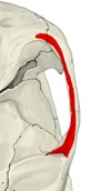 Origin of masseter
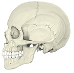 Skull (left lateral view)
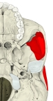 Temporal fossa (from below)
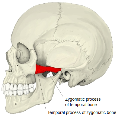
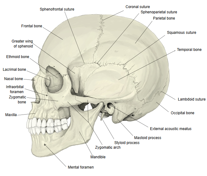
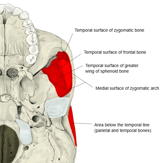
Other points
· Seen from the front, the skull is widest at the zygomatic arches.
Zygion: the most laterally projecting point of the zygomatic arch. The distance between the right and left zygia is taken as the facial (bizygomatic) breadth of the skull.
Behind, the upper border of the arch is continuous with the supramastoid crest of the temporal bone; the temporal fascia, which here splits into two layers, is attached to it.
The buccal fat pad lies in the hollow below the arch, between the masseter and the cheek. Unlike ordinary subcutaneous fat, it is soft, loosely lobulated fat with few fibres. It is well developed in infants, where it is called the sucking pad.
Reference websites
[Wikipedia]
· Zygomatic arch, Wikipedia.
· Temporal fossa, Wikipedia.
[Websites with illustrations]
· Zygomatic bone (Kenhub)
· Bones of the skull (TeachMeAnatomy)
Structure
Behind, the zygomatic process of the temporal bone arises by two roots:
· the anterior root runs medially in front of the mandibular fossa, where it widens into the articular tubercle;
· the posterior root runs backwards above the external acoustic meatus and continues as the supramastoid crest.
The upper border of the arch gives attachment to the temporal fascia; the lower border and medial surface give origin to the masseter. The arch bridges over the temporal fossa: the tendon of the temporalis passes deep to it to reach the coronoid process of the mandible. The arch can be fractured by a direct blow to the side of the face; a depressed fracture may press on the temporalis and make opening the mouth painful.
Where it is found
Related terms
- Temporal process
- Zygomatic process
- Masseter
- Temporal line
- Temporomandibular joint
- Zygomaticofacial foramen
- Zygomaticotemporal foramen
References and sources
- Further reading: Zygomatic arch, Wikipedia.
- Further reading: Temporal fossa, Wikipedia.
- Federative International Programme for Anatomical Terminology (FIPAT). Terminologia Anatomica, 2nd ed. (TA2), 2019.