Acinar lumen
Also called Lumen of acinus or the acinus lumen
- Overview
- Lumen and acinar cells
- Reference websites
- (Wikipedia, websites with illustrations)
- Related terms
Overview
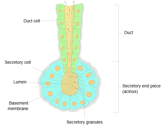
· The wall of the acinar lumen is formed directly by the acinar cells (their apical surfaces).
The figures below show, in simple form, the acini of the pancreas and the flow of pancreatic juice.
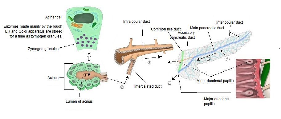
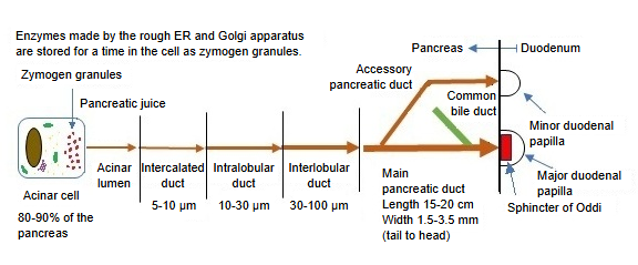
In the pancreas, the lumen often contains the pale centroacinar cells, the first cells of the intercalated duct pushed into the acinus. From the intercalated duct the juice passes through the intralobular and interlobular ducts into the main and accessory pancreatic ducts, and on into the duodenum.
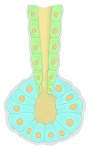 Exocrine gland (scheme)
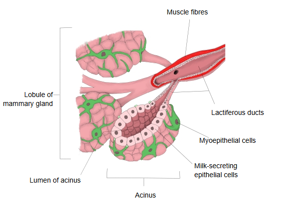
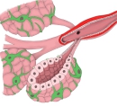 Acini of the mammary gland
Lumen and acinar cells
Does every acinus have a lumen?
In an acinus the acinar cells are arranged around the lumen, and the lumen is part of the structure that defines an acinus.
So it is fair to say that every acinus has a lumen. It is narrow in serous acini and wider in mucous glands and in the lactating mammary gland, where milk collects in it before passing into the lactiferous ducts.
Is there a membrane-like layer on the outside of the lumen, where it meets the acinar cells?
No. The lumen is bounded directly by the acinar cells; no separate membrane-like structure lies between the lumen and the cells.
Reference websites
[Wikipedia]
· Acinus, Wikipedia.
· Pancreatic acinar cell, Wikipedia.
[Websites with illustrations]
· Salivary gland acini and ducts (figure) (Pocket Dentistry)
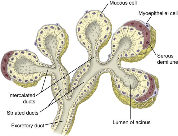
· Pancreatic acinus and ducts (figure) (DoctorLib)

· Pancreatic acinus with centroacinar cells (micrograph) (Wikimedia Commons)

Related terms
- Glandular cell
- Acinar cells
- Centroacinar cells
- Intercalated duct
- Glandular lumen
- Exocrine gland
- Pancreas
- Lobes of mammary gland
References and sources
- Further reading: Acinus, Wikipedia.
- Further reading: Pancreatic acinar cell, Wikipedia.
- Federative International Programme for Anatomical Terminology (FIPAT). Terminologia Anatomica, 2nd ed. (TA2), 2019.