Pancreas (Latin: pancreas)
- Overview
- Location
- Divisions
- Structure (exocrine part, endocrine part)
- Functions
- Vessels and nerves (arteries, veins, lymphatics, nerves)
- Related terms
- References
Overview
Location
[Position in the body]
The pancreas runs across the upper abdomen from its right lateral part through the middle to the left lateral part.
[Position relative to the skeleton]
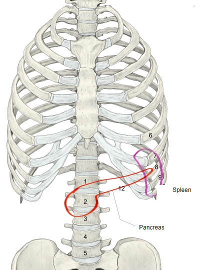
The head of the pancreas lies to the right of the vertebral bodies of the 1st–3rd lumbar vertebrae. The body lies in front of the vertebral column at the level of the 1st lumbar vertebra, and the tail ends near the spleen at the level of the 11th or 12th rib.
[Position relative to neighbouring organs]
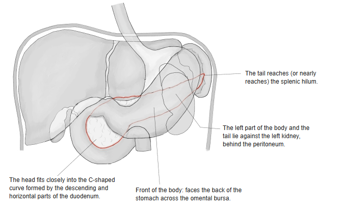
The pancreas is in contact with the great vessels of the abdomen. Behind the head lie the abdominal aorta, the inferior vena cava, the right renal vein and the lumbar part of the diaphragm. The superior mesenteric vein lies in the pancreatic notch, and the superior mesenteric artery often passes there too. The portal vein is formed near the upper border, and the gastroduodenal artery is seen in front of the head. Against the back of the body, the splenic artery runs along the upper border and the splenic vein below it, together with many lymph nodes and lymph vessels. The front of the body touches the stomach, and the root of the transverse mesocolon is attached to its anterior border.
The relations to the neighbouring organs are summarised below.
| Organ | Part of the pancreas | Relation | |
|---|---|---|---|
| 1 | Duodenum | Head | Surrounds it closely |
| 2 | Stomach | Body, neck | In contact in front (across the omental bursa) |
| 3 | Spleen | Tail | In contact (the tail reaches the splenic hilum) |
| 4 | Kidney (left) | Behind the body and tail | In contact retroperitoneally |
| 5 | Aorta | Behind the body and neck | Lies immediately behind |
| 6 | Common bile duct | Back of the head | Passes through it and often joins the pancreatic duct |
| 7 | Small intestine | Below the head | Touches the underside of the head |
| 8 | Transverse colon | In front of and below the body | In contact through its mesentery |
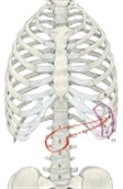 The pancreas and the vertebral column
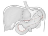 The pancreas and other organs 1
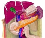 The pancreas and other organs 2
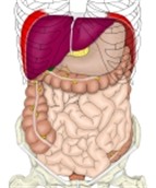 Organs in the abdominal cavity
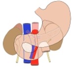 The pancreas and the great vessels
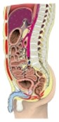 Trunk (sagittal section)
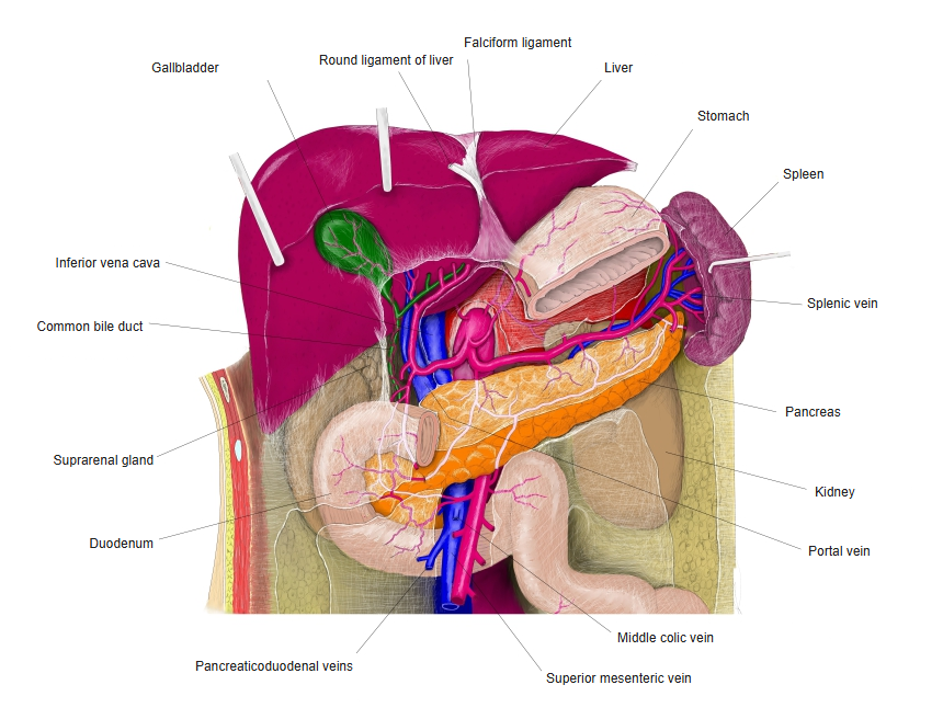
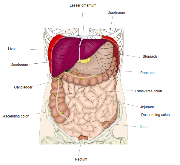
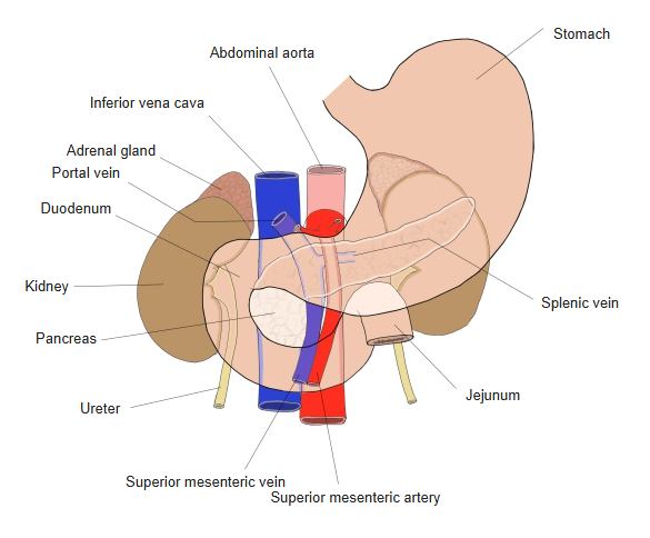
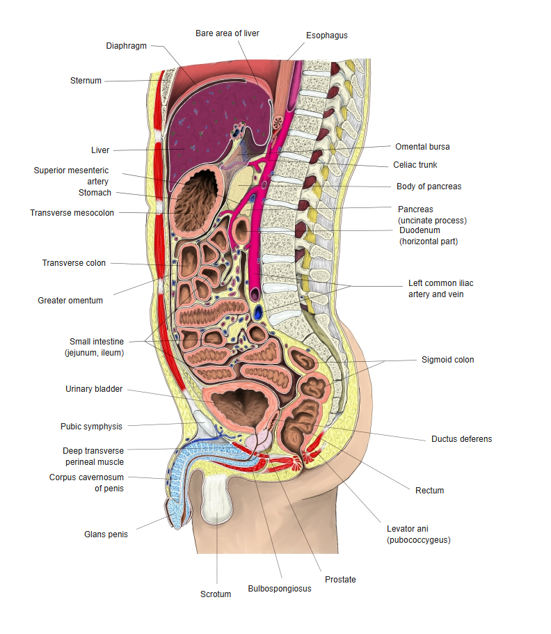
Divisions
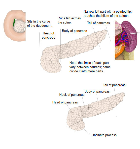
The pancreas can be divided into 3 parts: the head, the body and the tail.
the
pancreas
- Head : the swollen right end, fitting into the C-shaped curve of the duodenum. Related term: uncinate process.
- Body : runs to the left across the front of the vertebral column; it has 3 surfaces and 3 borders.
- Anterior surface : faces forwards and upwards and touches the back of the stomach across the omental bursa; covered by parietal peritoneum.
- Posterior surface : flat; joined directly to the posterior abdominal wall by loose connective tissue; not covered by peritoneum.
- Inferior surface : very narrow and faces forwards and downwards; sometimes absent; covered by parietal peritoneum.
- Anterior / superior / inferior borders : the anterior border is slightly sharp and gives attachment to the transverse mesocolon; the superior and inferior borders are blunt.
- Tail : the thin part extending furthest to the left; its tip is pointed and reaches about the middle of the visceral surface of the spleen.
Note: some sources divide the pancreas into 5 parts by adding 2 more (the neck and the uncinate process) to the 3 above.
Structure
structure of
the pancreas
- Exocrine part
- Acini : made of acinar cells; proportion 80–85%.
- Ducts : made of epithelial cells and centroacinar cells; proportion 5–10%.
- Endocrine part : 5 kinds of cells (β, α, δ, F/PP and ε cells) form the pancreatic islets (islets of Langerhans) and secrete hormones such as insulin and glucagon; proportion 1–2%.
- Others : connective tissue, blood vessels, nerves, stroma, immune cells and so on; proportion less than 1–3%.
The pancreas has a structure similar to that of the parotid gland. The connective tissue covering its surface enters the organ and divides the parenchyma into many lobules; the arteries, veins, lymph vessels and ducts run in the interlobular connective tissue. The lobules of the pancreas consist of an exocrine part and an endocrine part.
structure of
the pancreas
- Exocrine part (about 95% of the pancreas)
- Acini
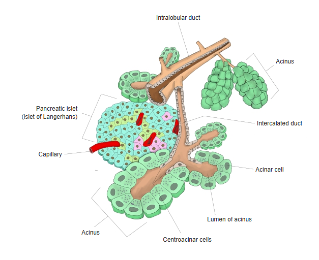
- Cuboidal / pyramidal acinar cells, forming a compound acinar gland, produce pancreatic juice.
- The acinar cells contain secretory granules (zymogen granules).
- Intercellular secretory canaliculi are seen between the cells.
- Flat centroacinar cells are seen on the inner surface of the centre of the acinus.
- Duct system : carries the pancreatic juice made in the acini to the pancreatic duct (cells: duct cells, centroacinar cells).
Pancreatic juice → acinar lumen → intercalated duct → intralobular duct → interlobular duct → main pancreatic duct
- Acini
- Endocrine part (about 1–2%) : Pancreatic islets
- Groups of endocrine cells scattered among the lobules (roughly round, surrounded by thin connective tissue).
- Particularly numerous in the tail; they have no ducts.
- Cells smaller than the acinar cells (about 15–16 µm across); about 1 million islets in all, each with about 1,000–4,000 cells.
- Cells: β cells (about 60–70%: insulin), α cells (about 15–20%: glucagon), δ cells (about 5–10%: somatostatin), PP cells (a few %: pancreatic polypeptide), ε cells (very few: ghrelin).
- The hormones made by each cell enter the capillaries in the islet and leave the pancreas.
- Cells of the pancreas with work other than secretion: fibroblasts, pancreatic stellate cells, vascular endothelial cells, immune cells, nerve cells.
 Acini and pancreatic islets
 Acinar cells
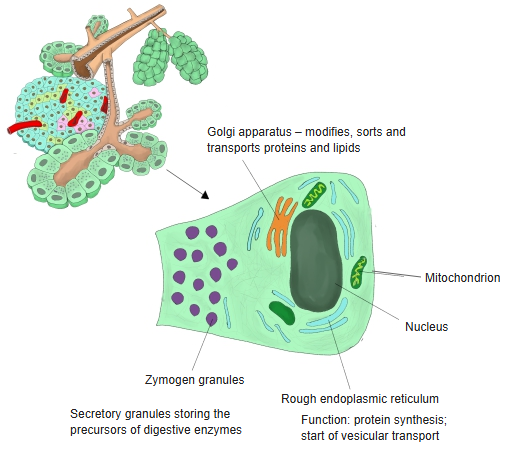
[Exocrine part]
structure of
the pancreas
- Exocrine part (about 95% of the pancreas)
- Acini
- Cuboidal / pyramidal acinar cells, forming a compound acinar gland, produce pancreatic juice.
- The acinar cells contain secretory granules (zymogen granules).
- Intercellular secretory canaliculi are seen between the cells.
- Flat centroacinar cells are seen on the inner surface of the centre of the acinus.
- Duct system : carries the pancreatic juice made in the acini to the pancreatic duct (cells: duct cells, centroacinar cells).
Pancreatic juice → acinar lumen → intercalated duct → intralobular duct → interlobular duct → main pancreatic duct
- Acini
- Endocrine part (about 1–2%) : pancreatic islets (see below).
- Cells of the pancreas with work other than secretion: fibroblasts, pancreatic stellate cells, vascular endothelial cells, immune cells, nerve cells.
- Acini and pancreatic islets
- Acinar cells
· Taking the exocrine part as 100, the ducts make up 2–5 and the rest is acinar cells.
[Other cells in the exocrine part]
The main cells of the exocrine tissue are the acinar cells and the duct epithelial cells, but the following cells are also found around them.
Myoepithelial cells: generally hardly seen in the pancreas; present in the salivary glands and elsewhere, but said to be almost absent from the exocrine pancreas.
Stromal cells: fibroblasts, vascular endothelial cells, immune cells and so on in the stroma (connective tissue) of the exocrine pancreas. They are not cells of the acini or ducts, but take part in supporting the tissue and supplying blood.
Immune cells (macrophages and others).
[Endocrine part]
structure of
the pancreas
- Exocrine part
- Acini : made of acinar cells; proportion 80–85%.
- Ducts : made of epithelial cells and centroacinar cells; proportion 5–10%.
- Endocrine part : pancreatic islets or islets of Langerhans; secrete hormones; proportion 1–2%.
- β cells : read "beta"; secrete insulin; proportion 60–70%.
- α cells : read "alpha"; secrete glucagon; proportion 15–20%.
- δ cells : read "delta"; secrete somatostatin; proportion 5–10%.
- F cells : (PP cells); secrete pancreatic polypeptide; proportion 1–5%.
- ε cells : read "epsilon"; secrete ghrelin; proportion under 1%.
- Others : connective tissue, blood vessels, nerves, stroma, immune cells and so on; proportion less than 1–3%.
| Name | Proportion (approx.) | Secretes | Action | |
|---|---|---|---|---|
| 1 | β cells (beta) | 60–70% | Insulin | Lowers blood glucose (promotes uptake of glucose into cells) |
| Size: about 10–20 µm; shape: polygonal to slightly rounded; other: central nucleus, many secretory granules | ||||
| 2 | α cells (alpha) | 15–20% | Glucagon | Raises blood glucose (promotes breakdown of glycogen in the liver) |
| Size: about the same as β cells; shape: slightly oval; other: more numerous at the edge of the islet than β cells | ||||
| 3 | δ cells (delta) | 5–10% | Somatostatin | Inhibits the secretion of insulin and glucagon |
| Size: slightly smaller; shape: irregular; other: granules relatively small and dense | ||||
| 4 | F cells (PP cells) | 1–5% | Pancreatic polypeptide | Regulates the movement of the gastrointestinal tract and the secretion of pancreatic juice |
| Size: small; shape: irregular; other: a few at the edge of the islets; secretory granules fine and scattered | ||||
| 5 | ε cells (epsilon) | Very few | Ghrelin | Appetite-stimulating hormone |
| Size: smallest; shape: irregular to slightly spindle-shaped; other: very few; many in the fetus, very few in adults | ||||
- Acini and pancreatic islets
- Acinar cells
Functions
[Helping to digest food]
The acinar cells, which make up most of the pancreas (about 80–85%), secrete pancreatic juice containing digestive enzymes, which work as follows.
| Name | Notes | |
|---|---|---|
| 1 | Amylase | Breaks down starch to the disaccharide maltose. |
| 2 | Lipase | Breaks down fats into fatty acids and monoglycerides. |
| 3 | (Chymo)trypsin | Breaks down proteins (peptones) into peptides and amino acids. |
| 4 | Nucleases | Break down nucleic acids. |
In addition, the bicarbonate ions in the pancreatic juice (and in the bile) neutralise gastric acid and keep the contents of the small intestine alkaline.
[Regulating blood glucose]
The cells in the pancreatic islets (islets of Langerhans) secrete hormones that regulate blood glucose.
| Name | Proportion (approx.) | Secretes | Action | |
|---|---|---|---|---|
| 1 | β cells (beta) | 60–70% | Insulin | Lowers blood glucose |
| 2 | α cells (alpha) | 15–20% | Glucagon | Raises blood glucose |
| 3 | δ cells (delta) | 5–10% | Somatostatin | Inhibits the secretion of insulin and glucagon |
| 4 | F cells (PP cells) | 1–5% | Pancreatic polypeptide | Regulates gastrointestinal movement and the secretion of pancreatic juice |
| 5 | ε cells (epsilon) | Very few | Ghrelin | Appetite-stimulating hormone |
Vessels and nerves
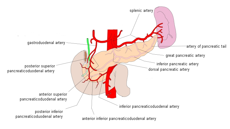
[Arteries]
· Head: front) anterior superior pancreaticoduodenal artery, anterior inferior pancreaticoduodenal artery; back) posterior superior pancreaticoduodenal artery, posterior inferior pancreaticoduodenal artery.
· Body: the dorsal pancreatic artery, inferior pancreatic artery and great pancreatic artery, branches of the splenic artery.
· Tail: the artery of the pancreatic tail, a branch of the splenic artery.
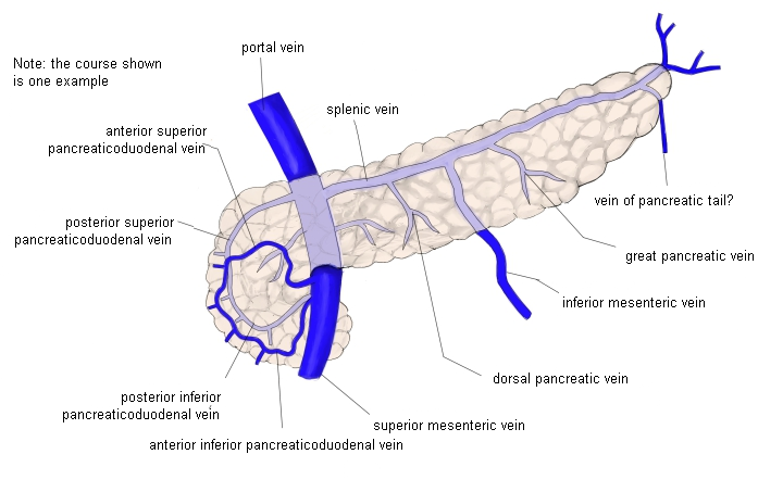
[Veins]
There are few sources on the veins, but basically it seems right to think that "companion veins with the same names as the arteries collect the blood from the pancreas".
· Head: companion veins of the arteries are present.
The anterior superior pancreaticoduodenal vein and posterior superior pancreaticoduodenal vein drain into the superior mesenteric vein, either separately or after joining into one (superior pancreaticoduodenal vein).
The posterior inferior pancreaticoduodenal vein and anterior inferior pancreaticoduodenal vein join (inferior pancreaticoduodenal vein) and drain into the superior mesenteric vein.
· Body / tail: the dorsal pancreatic vein, inferior pancreatic vein, great pancreatic vein and vein of the pancreatic tail collect the venous blood of the body and tail and drain into the splenic vein.
The pancreaticoduodenal veins accompany the superior and inferior pancreaticoduodenal arteries to the portal vein, and the pancreatic veins enter the splenic vein behind.
The veins of the pancreas flow mainly into the splenic vein and the superior mesenteric vein, which join to form the portal vein.
Blood is sent to the liver through the portal vein.
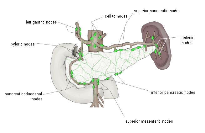
[Lymphatics]
The pancreas produces large amounts of digestive enzymes as an exocrine gland and also has endocrine functions, so a rich network of lymph vessels, involved in metabolism and immune responses, spreads through it.
[Features of the lymph vessels of the pancreas]
Lymph vessels within the parenchyma
· Fine lymph capillaries are distributed around the acini and islets.
Interlobular lymph vessels
· Run in the interlobular connective tissue alongside the vessels and ducts.
Large lymph vessels
· Flow into the superior mesenteric, splenic hilar and celiac lymph nodes and others.
· Head → mainly the superior pancreaticoduodenal nodes.
· Body and tail → the splenic hilar nodes and celiac nodes.
· The lymph of the pancreas flows into the lymph nodes around it.
· The main nodes are the pancreaticoduodenal nodes, the splenic nodes and the superior mesenteric nodes.
· From these it flows on to the celiac nodes and finally into the thoracic duct.
[Nerves]
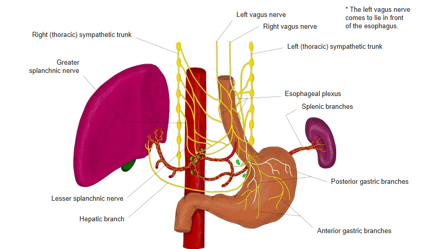
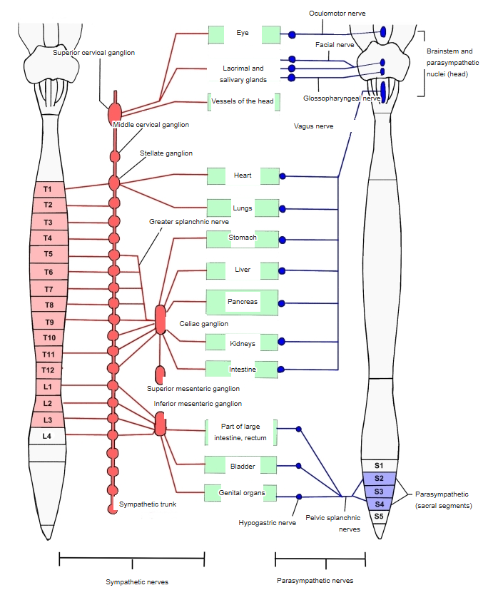
The pancreas is supplied mainly by the autonomic nervous system, and the nerves related to it basically pass through the pancreatic plexus.
The autonomic nerve fibres distributed to the pancreas (sympathetic, parasympathetic and afferent) in principle all reach it through the pancreatic plexus; the pancreatic plexus acts as a kind of hub.
[Sympathetic nerves]
Sympathetic fibres, mainly from the thoracic sympathetic ganglia, enter the pancreas through the celiac plexus and the superior mesenteric plexus.
They constrict the blood vessels of the pancreas and inhibit the secretion of pancreatic juice and hormones.
· The pancreatic plexus spreads over the surface and through the whole parenchyma; parasympathetic and afferent fibres also join it.
· The sympathetic fibres synapse in the celiac and superior mesenteric ganglia, so they pass through the pancreatic plexus as postganglionic fibres; the ganglion cells in the pancreatic plexus are where the parasympathetic (vagal) fibres synapse.
[Parasympathetic nerves]
The pancreas is supplied mainly by the vagus nerve (cranial nerve X).
It promotes the secretion of pancreatic juice and of insulin.
Parasympathetic fibres also reach the pancreas through the celiac plexus.
Unmyelinated nerve fibres from the sympathetic celiac plexus are distributed directly, or enter the pancreas together with fibres from the hepatic plexus and the splenic plexus.
The parasympathetic vagus nerve enters the pancreas from the stomach wall through the walls of the pylorus and duodenum. The secretory fibres are said to run along the superior pancreaticoduodenal artery and vein. In the interlobular connective tissue, sympathetic ganglion cells are found, and lamellated corpuscles are found as endings of sensory nerves.
nerve
- Left vagus nerve (anterior vagal trunk) : terminal branch.
- Right vagus nerve (posterior vagal trunk) : terminal branch ⇒ Celiac branch ⇒ celiac plexus ⇒ renal, pancreatic and intestinal branches
- The pancreatic branches help form the pancreatic plexus, which contains not only parasympathetic fibres from the vagus nerve but also sympathetic and afferent fibres.
· The vagus nerves enter the abdominal cavity through the esophageal hiatus of the diaphragm and divide into left and right terminal branches.
· In the pancreas the vagus nerve influences both endocrine and exocrine secretion.
[Visceral afferent fibres (sensory fibres)]
· They run along the autonomic nerve fibres and carry stretch of the pancreas, chemical stimuli, inflammation and pain (dull visceral pain) to the brain.
· They travel mainly by the vagus nerve and by the sympathetic route (to the dorsal root ganglia) (afferent nerve fibres).
 Vagus nerve (thorax and abdomen)
 Autonomic nervous system
Related terms
Main anatomical terms related to the pancreas, in alphabetical order.
References and sources
- Further reading: Pancreas, Wikipedia.