Acinus of mammary gland (Latin: acinus glandulae mammariae)
Also called: alveolus of mammary gland
· Anatomy textbooks say that the glandular membrane of the acinus has 'basket cells'. Here this is an older name for the myoepithelial cells, which embrace the acinus like a basket; it is not the basket cell of the cerebellum.
· The size and shape of the acini vary greatly with the individual, age and functional state.
· During milk secretion the glandular cells are very active: large fat droplets are released into the lumen by apocrine secretion as milk fat globules, while the milk proteins leave the cells by exocytosis.
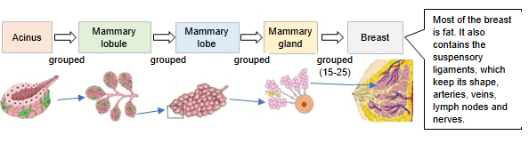
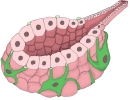 Acinus (section)
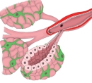 Mammary lobule
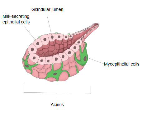
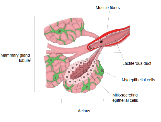
Anatomy textbooks describe the acini of the mammary gland as follows.
Each lobe of the mammary gland is divided into mammary lobules filled with acini (the secretory end-pieces); the spaces between the lobes and lobules are filled with connective tissue and fat. The size and shape of the acini vary greatly with the individual, age and functional state. Before any pregnancy the acini are very small and filled with glandular cells, so that no lumen can be seen yet. In pregnancy most acini are completed, and in lactation they become fully functional. The acinus consists of a glandular membrane with basket (myoepithelial) cells and an epithelium of glandular cells, and the width of its lumen depends on the amount secreted. During lactation the fat is released into the lumen by apocrine secretion as milk fat globules; in the early phase the secretion also contains colostrum corpuscles.
Reference websites
[Wikipedia]
· Mammary gland, Wikipedia.
[Websites with illustrations]
· Breast and mammary gland with acini, milk-secreting and myoepithelial cells (figure) (Semantic Scholar (figure))

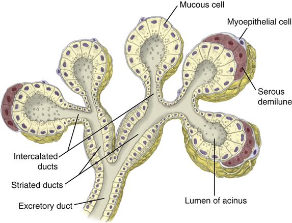
· Glandular acini and ducts (salivary gland, for comparison) (Pocket Dentistry)
Related terms
- Lobule of mammary gland
- Lobes of mammary gland
- Myoepithelial cell
- Lactiferous duct
- Acinar lumen
- Mammary gland
- Acinus (glandular acinus)
References and sources
- Further reading: Mammary gland, Wikipedia.
- Federative International Programme for Anatomical Terminology (FIPAT). Terminologia Anatomica, 2nd ed. (TA2), 2019.