Cardiac muscle (Latin: textus muscularis striatus cardiacus)
Also called the heart muscle or the cardiac muscle tissue
The muscle that makes up the heart: the striated, involuntary muscle tissue that forms the myocardium, the thick muscle layer of the heart wall.
More detail
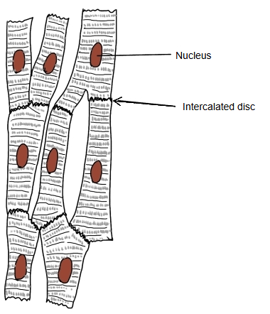
Like skeletal muscle, cardiac muscle is a striated muscle; but whereas skeletal muscle is a multinucleate voluntary muscle, cardiac muscle is an involuntary muscle whose cells (cardiomyocytes) usually have one central nucleus (rarely two). Like smooth muscle it is involuntary: its beat starts on its own in the conducting system of the heart, and autonomic nerves only speed it up or slow it down. The cells are very rich in mitochondria, which supply the energy (ATP) for a heart that contracts without rest throughout life.
The cells of cardiac muscle are short and branched and are joined into a network; the junctions between them are called intercalated discs. Intercalated discs play an important part in passing the electrical signal of the heart: their gap junctions let the signal pass quickly from cell to cell, so the heart muscle contracts as one unit, and their desmosomes hold the cells together.
A chain of cardiac muscle cells joined end to end is called a cardiac muscle fiber. Adult cardiac muscle cells divide very little, so lost heart muscle is replaced mainly by scar tissue.
Reference websites
[Wikipedia]
· Cardiac muscle, Wikipedia.
· Intercalated disc, Wikipedia.
Related terms
- Myocardium
- Heart
- Striated muscle
- Skeletal muscle
- Smooth muscle
- Involuntary muscle
- Conducting system of the heart
References and sources
- Further reading: Cardiac muscle, Wikipedia.
- Further reading: Intercalated disc, Wikipedia.
- Federative International Programme for Anatomical Terminology (FIPAT). Terminologia Anatomica, 2nd ed. (TA2), 2019.