Circular folds (Latin: plicae circulares)
Also called the valves of Kerckring
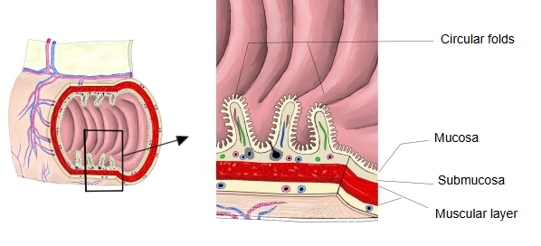
a circular fold
- Mucosa : covers the surface of the fold; made of the epithelium (simple columnar epithelium), the lamina propria and the muscularis mucosae.
- Submucosa : a fairly thick layer of connective tissue under the mucosa, with blood vessels and nerves.
· The circular folds of the jejunum are taller and more closely packed than those of any other part, which raises its absorption of nutrients.
Differences along the small intestine
| Feature | Duodenum | Jejunum | Ileum | |
|---|---|---|---|---|
| 1 | Development | Moderate (absent from the duodenal bulb; clear beyond it) | Best developed | Become fewer; almost absent at the end |
| 2 | Height | Low to medium | Tall and large (up to about 8–10 mm) | Low and shallow |
| 3 | Density | Medium | Very dense | Sparse |
| 4 | Distribution | Well developed in the descending and horizontal parts | Densely packed in the upper and middle small intestine | Sparser towards the ileocaecal region |
| 5 | Functional meaning | Mixing with the digestive juices | The main site of absorption | Absorbs, but mainly reabsorbs bile acids (bile salts) and vitamin B12 |
| 6 | Shape | Somewhat irregular, spiral | Regular, tall rings | Low, tending to be discontinuous |
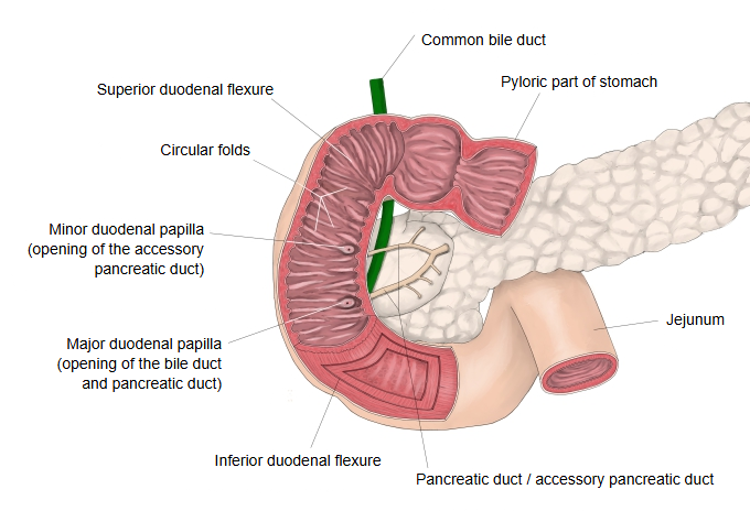
Unlike the rugae of the stomach, the circular folds do not flatten out when the intestine is distended. They enlarge the absorptive surface about threefold and slow the passage of chyme, giving more time for digestion and absorption; their surface is in turn covered with villi, and the villi with microvilli. On imaging they cross the full width of the bowel, which helps to tell small intestine from colon (whose haustral folds do not cross the whole lumen). The major duodenal papilla lies on a fold of the descending part of the duodenum.
Folds compared along the digestive tract
Where it is found
Related terms
References and sources
- Further reading: Circular folds, Wikipedia.
- Further reading: Small intestine, Wikipedia.
- Federative International Programme for Anatomical Terminology (FIPAT). Terminologia Anatomica, 2nd ed. (TA2), 2019.