Jejunum (Latin: jejunum)
- Overview
- Relations to other organs
- Structure of the jejunal wall (mucosa [epithelium, lamina propria, muscularis mucosae], submucosa, muscle layer, subserosa, serosa)
- Functions
- Vessels and nerves (arteries, veins, lymphatics, nerves)
- Jejunum and ileum compared (diameters, absorption, summary)
- Related terms
- References
Overview
- Respiratory system
- Digestive system : oral cavity, pharynx, esophagus, stomach, small intestine, large intestine
- Urinary system
- Genital system
- Peritoneum
- Endocrine glands
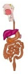
· The length of the jejunum and ileum differs greatly between the living body and the cadaver: about 2–3 m in life and up to 6–7 m after death, mainly because muscle tone is lost and the intestine is stretched by gravity and handling.
· In their natural winding course in the abdominal cavity, the jejunum lies mostly in the umbilical region, the lower abdomen and the left iliac fossa, and the ileum in the greater pelvis, the lower abdomen and the right iliac fossa.
small intestine
- Duodenum : C-shaped tube (about 25–30 cm) continuing from the stomach; receives pancreatic juice and bile.
- Jejunum : continues from the duodenum and forms the upper 2/5 of the small intestine beyond it; the main site of absorption of the (three major) nutrients.
- Ileum : the last part of the small intestine (the lower 3/5); absorbs the nutrients not absorbed in the jejunum.
Relations to other organs
The jejunum winds fairly freely in the abdominal cavity. It is attached to the posterior abdominal wall by the mesentery but remains mobile, so the organs it touches vary between individuals and with how full the intestine is. In general they are as follows.
Organs the jejunum commonly touches
1. Ileum: continuous with the jejunum; the border between them is not distinct.
2. Transverse colon and transverse mesocolon: they lie above the jejunum, so the proximal jejunum in particular may touch them.
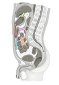 Abdominal and pelvic cavities
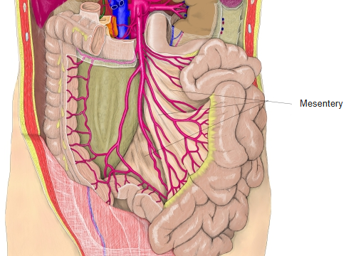
(longitudinal section)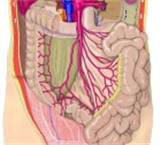 Arteries of the
abdominal cavity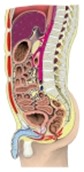 Trunk
(sagittal section)
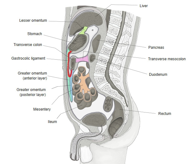
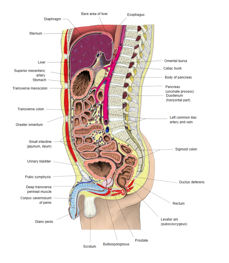
3. Ascending colon: loops on the right of the jejunum may touch it, although the ileum is generally in closer contact.
4. Descending colon: lies in the left flank, so loops on the left of the jejunum may touch it.
5. Stomach (especially the pyloric part and the greater curvature): the beginning of the jejunum lies below the stomach, so it may come close to it indirectly.
6. Pancreas (near the head and the uncinate process): close to it by way of the duodenum.
7. Greater and lesser omentum: they often cover the jejunum from above and in front.
8. Abdominal wall and retroperitoneal structures (kidneys, ureters and others): close to them through the mesentery rather than in direct contact.
In short, besides the ileum the jejunum may touch the transverse colon, the ascending and descending colon, the stomach and the greater omentum, but these contacts are not fixed: they change with intestinal movement and filling.
Structure of the jejunal wall
The following summarizes the structure of the jejunal wall.
jejunal wall
- Mucosa
- Epithelium : almost entirely villi and crypts; flat areas are very few. Cells: enterocytes, goblet cells, enteroendocrine cells, Paneth cells, stem cells.
- Lamina propria : fills the villi and surrounds the crypts. Contents: cells (fibroblasts and others), fibres, blood vessels, lymph vessels, nerves and others.
- Muscularis mucosae : a thin layer of smooth muscle with inner circular and outer longitudinal parts.
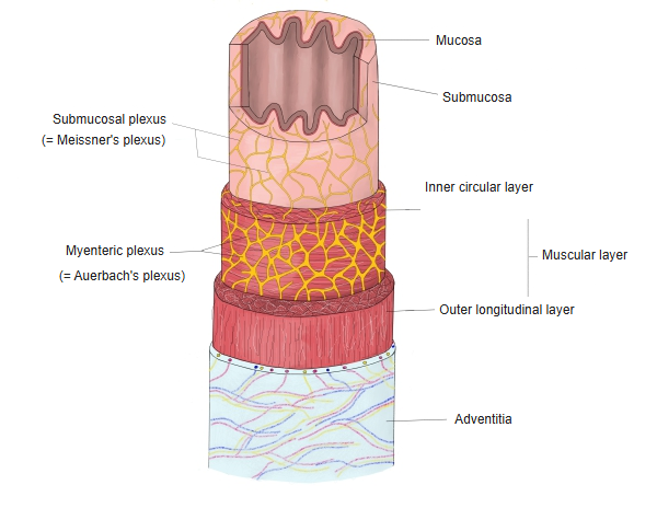
- Submucosa : flexible loose connective tissue. Other contents: blood vessels, lymph vessels, a nerve plexus (submucosal plexus = Meissner's plexus), immune tissue and others.
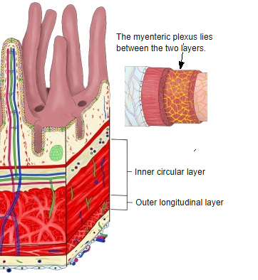
- Muscle layer
- Inner circular layer : smooth muscle running around the tube; thicker than the outer longitudinal layer.
- Outer longitudinal layer : smooth muscle running along the tube.
- Subserosa : loose connective tissue with blood vessels, nerves and lymph vessels.
- Serosa : two layers: an inner thin connective tissue layer (with vessels and nerves) and an outer simple squamous epithelium (which secretes serous fluid).
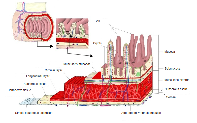
[Mucosa]
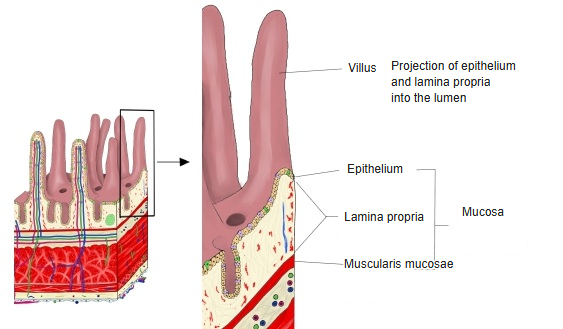
Epithelium
- Villi : projections towards the lumen, 0.5–1.2 mm long, most of them arising from the circular folds. Functions: absorption of nutrients, mucus and hormone secretion. Cells: enterocytes, goblet cells, enteroendocrine cells.
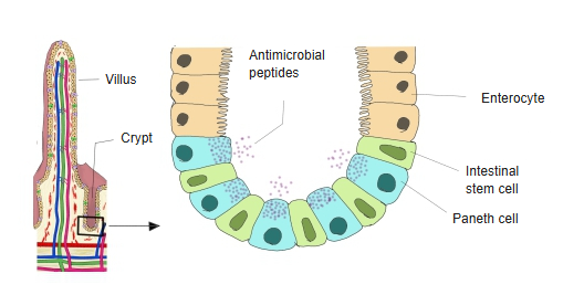
- Crypts : tubular pits at the bases of the villi, 100–200 µm deep. Functions: supplying and maintaining epithelial cells, defence. Cells: stem cells, Paneth cells, differentiating cells.
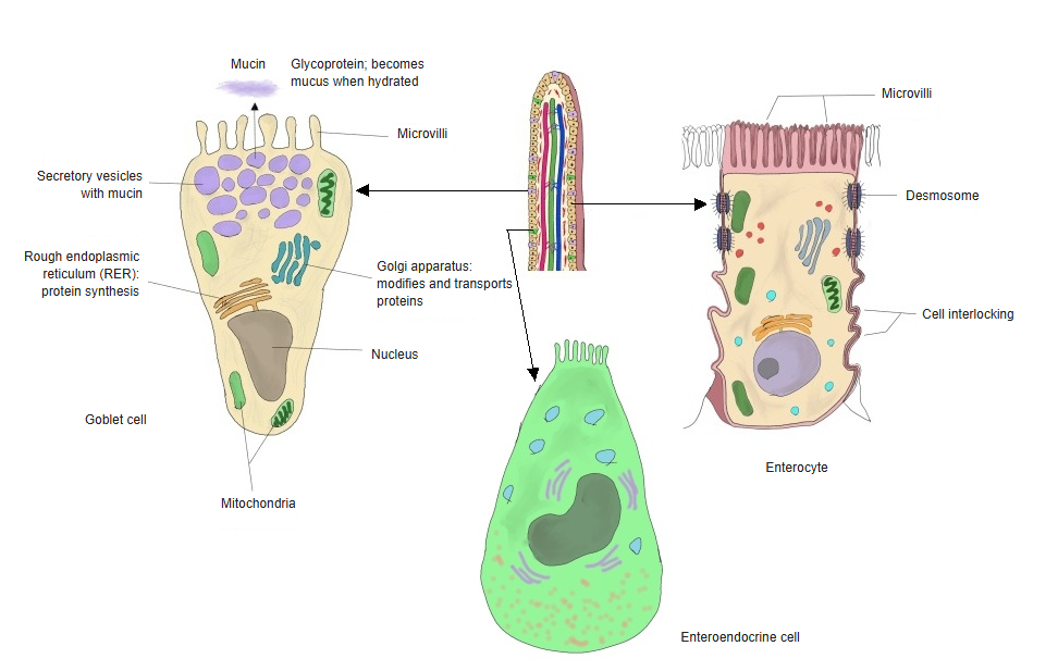
The epithelium is made of the following five kinds of cells.
| Name | Proportion (%) | Notes | |
| 1 | Enterocytes | 80–90 | Have microvilli on their surface; the main cells for absorbing nutrients. |
| 2 | Goblet cells | 5–15 | Secrete mucus; they increase in number further down the intestine. |
| 3 | Enteroendocrine cells | 1–2 | Secrete gastrointestinal hormones (serotonin, cholecystokinin, secretin and others). |
| 4 | Paneth cells | 1–5 | At the bottom of the crypts; secrete antimicrobial substances. |
| 5 | Stem cells | a few | Divide and differentiate, constantly supplying new epithelial cells. |
· The proportions are approximate and differ somewhat between sources.
 Cells of the villi
 Bottom of a crypt
Lamina propria
The lamina propria is made of the following components.
| Name | Notes | |
| 1 | Cells | Fibroblasts, macrophages, dendritic cells, plasma cells, endothelial cells (forming blood and lymph vessels). |
| 2 | Extracellular components | Collagen fibres (mainly types I and III), elastic fibres, ground substance (proteoglycans, hyaluronic acid and others). |
| 3 | Blood and lymph vessels | Central lacteal, capillary network. |
| 4 | Immune structures | Lymphocytes; connected with the lymphoid follicles of the Peyer's patches (gut-associated lymphoid tissue, GALT); take part in the immune defence of the small intestine. |
Muscularis mucosae
· Its thickness and development differ between organs, but the muscularis mucosae is continuous from the esophagus to the rectum.
[Submucosa]
· Its nerves form a plexus (the submucosal plexus = Meissner's plexus).
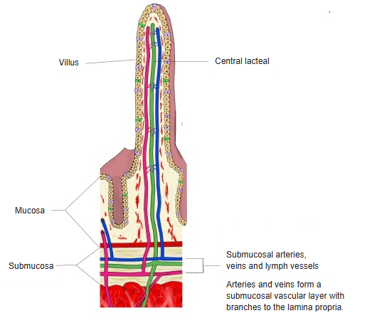
Its components are summarized below.
| Name | Notes | |
| 1 | Blood vessels | Supply oxygen and nutrients to the mucosa and carry absorbed nutrients to the portal system; they have no particular names. |
| 2 | Lymph vessels | A well-developed lymphatic plexus; lymph from the central lacteals collects here. |
| 3 | Nerve plexus | The submucosal plexus (Meissner's plexus) regulates secretion, local blood flow and movements of the mucosa. |
| 4 | Fibres | Collagen and elastic fibres, which keep the tissue elastic and strong. |
| 5 | Immune tissue | Solitary lymphoid nodules, Peyer's patches (less developed than in the ileum). |
 Submucosal plexus
(esophagus)
[Muscle layer]
- Inner circular layer : fibres running around the tube; thicker than the outer layer; helps to mix the contents and to produce peristalsis.
- Outer longitudinal layer : fibres running along the tube; helps to drive peristalsis forward.
· The thickness of the muscle layer changes greatly with its state of contraction (roughly 0.2–0.5 mm).
The muscle layer along the digestive tract
| Part | Features | |
| 1 | Esophagus | Upper third striated (skeletal) muscle, middle third striated and smooth, lower third smooth muscle; inner circular and outer longitudinal layers throughout. |
| 2 | Stomach | Three layers (outer longitudinal, middle circular, inner oblique). |
| 3 | Small intestine (jejunum and ileum) | The standard two layers; the inner circular layer is a little thicker, and peristalsis is active. |
| 4 | Large intestine | Two layers, but the outer longitudinal muscle is gathered into three bands (taeniae coli). |
| 5 | Rectum and anal canal | From the lower rectum to the anal sphincters, skeletal muscle is mixed in (external anal sphincter). |
[Subserosa]
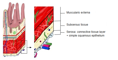
| Name | Notes | |
| 1 | Loose connective tissue | Loose connective tissue with collagen and elastic fibres, binding the serosa loosely to the muscle layer beneath. |
| 2 | Blood vessels | Small arteries and veins that supply the serosa and the muscle layer of the small intestine; they have no particular names. |
| 3 | Lymph vessels | Part of the lymph drainage of the small intestine, especially after fat absorption. |
| 4 | Adipose tissue | Tends to be more abundant in the ileum than in the jejunum; varies between individuals and with age and nutrition. |
| 5 | Nerve plexus | Subserous plexus: part of the autonomic nerves of the intestine, regulating the muscle layer and the blood vessels. |
[Serosa]
- Mesothelium : simple squamous epithelium facing the peritoneal cavity; secretes serous fluid.
- Connective tissue : thin loose connective tissue with small blood vessels, lymph vessels and nerves.
Functions
Its main functions are as follows.
Absorption of nutrients (the main function)
1. Carbohydrates are absorbed as monosaccharides (glucose, galactose, fructose).
2. Proteins are absorbed as amino acids and dipeptides.
3. Fats are absorbed as fatty acids and monoglycerides (emulsification → micelle formation → into the central lacteals).
4. Vitamins and minerals (water-soluble and fat-soluble vitamins, iron, calcium and others) are also absorbed mainly here.
Absorption of water and electrolytes
Much of the water in the digestive juices mixed with the food is absorbed in the jejunum.
Final stage of digestion
Besides the digestive enzymes coming from the duodenum, the brush-border enzymes of the jejunum break sugars and peptides down further into forms that are easy to absorb.
| Jejunum: the main site of digestion and absorption | Ileum: the rest, and special substances |
Mainly absorbed in the jejunum: · Sugars (glucose, galactose and others) · Amino acids, peptides · Fatty acids, monoglycerides · Vitamins (water-soluble and fat-soluble) · Water and electrolytes (water absorption is most active in the jejunum) · Others: iron, calcium | Mainly absorbed in the ileum: · Vitamin B12 (absorbed only in the terminal ileum, as a complex with intrinsic factor) · Bile acids (reabsorbed in the terminal ileum and returned to the liver) · The remaining fats · Water and electrolytes (sodium absorption somewhat predominant) |
Vessels and nerves
[Arteries]
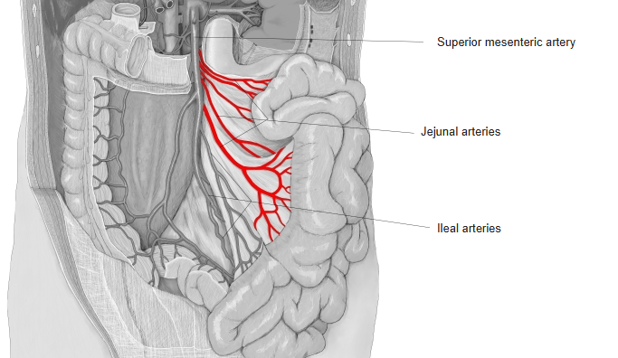
The jejunal arteries, branches of the superior mesenteric artery, form arterial arcades near the jejunum, from which straight arteries (vasa recta) pass to the jejunum.
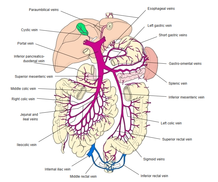
[Veins]
The jejunal veins accompany the jejunal arteries.
Jejunum ⇒ straight veins ⇒ venous arcades ⇒ jejunal veins ⇒ superior mesenteric vein (the venous arcades are less distinct than the arterial ones and vary more).
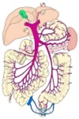 Superior mesenteric vein
[Lymphatics]
The lymph flow in the jejunum is as follows.
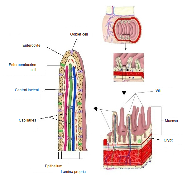
1. Lymph begins in the central lacteal that runs in the centre of each villus (part of the mucosa). Fat absorbed by the villi passes into the central lacteals, and the fat-rich lymph looks milky; it is called chyle.
2. From the central lacteals the lymph passes through the submucosa, the muscle layer and the serosa.
3. Finally it leaves the serosa and enters the mesentery.
The flow of lymph after it leaves the intestine is summarized below.
[Nerves]
The jejunum is controlled mainly by the enteric plexuses of the autonomic nervous system, which also receive input from the rest of the autonomic nervous system.
· The enteric plexuses consist of two parts:
1. Submucosal plexus (Meissner's plexus)
2. Myenteric plexus (Auerbach's plexus)
- Mucosa : epithelium, lamina propria, muscularis mucosae.
- Submucosa : submucosal plexus (Meissner's plexus) ⇒ enteric plexuses (autonomic), which control the whole digestive tract.
- Muscle layer : inner circular and outer longitudinal layers, with the myenteric plexus (Auerbach's plexus) between them ⇒ enteric plexuses.
- Subserosa
- Serosa
The input from the autonomic nervous system is as follows.
1. Sympathetic: from the superior mesenteric plexus → inhibits peristalsis and constricts the vessels.
2. Parasympathetic: the vagus nerve → promotes peristalsis and secretion.
to the jejunum
- Sympathetic fibres : from the sympathetic trunk (T5–T12) through the superior mesenteric plexus, along the jejunal and ileal arteries. Actions: inhibit peristalsis, constrict vessels, inhibit secretion.
- Parasympathetic fibres : from the vagus nerve (CN X). Actions: promote peristalsis and secretion, increase blood flow.
- Sensory fibres : visceral afferent fibres. Action: sense mechanical and chemical stimuli in the mucosa and convey them to the centre.
Jejunum and ileum compared
[Diameters]
| Part | Outer diameter (cm) | Inner diameter (cm) | Notes | |
| Jejunum | Upper (proximal) | about 3.0–3.5 | about 2.5–3.0 | The widest part; the circular folds are well developed. |
| Lower (distal) | about 2.8–3.2 | about 2.3–2.8 | Gradually narrower. | |
| Ileum | Middle | about 2.5–2.8 | about 2.0–2.5 | Lymphoid tissue (Peyer's patches) increases. |
| Terminal | about 2.0–2.5 | about 1.5–2.0 | The narrowest part; continues to the ileocecal valve. | |
· The values are approximate.
[Absorption]
| Item | Jejunum | Ileum | |
| 1 | Main role | Main absorption of nutrients | Absorption of special substances + the rest |
| 2 | Efficiency | Very high | Lower than in the jejunum |
| 3 | Substances absorbed | Sugars, amino acids, fats, water, electrolytes, Ca, Fe | Vitamin B12, bile acids, Na, the remaining nutrients |
| 4 | Tissue features | Tall villi, many folds | Lower villi, many Peyer's patches |
| 5 | Transport mechanisms | Varied and numerous | Special transporters (vitamin B12, bile acids) |
[Summary]
| Item | Jejunum | Ileum | |
| 1 | Position | Upper part of the small intestine, continuing from the duodenum | Lower part of the small intestine, continuing from the jejunum to the cecum |
| 2 | Length | About 2/5 of the jejunum and ileum | About 3/5 |
| 3 | Wall | Thick | Thin |
| 4 | Blood vessels | Rich (larger vessels forming a few arcades, long vasa recta) | Fewer (smaller vessels forming many arcades, short vasa recta) |
| 5 | Fat | Little fat in the mesentery | Much fat in the mesentery, reaching close to the intestine |
| 6 | Folds | High, dense and well developed | Low and sparse; almost absent at the end |
| 7 | Lymphoid tissue | Mainly solitary lymphoid nodules | Many Peyer's patches (aggregated lymphoid nodules) |
| 8 | Colour | Redder | Pale pink |
Related terms
Main anatomical terms related to the jejunum, in alphabetical order.
References and sources
- Further reading: Jejunum, Wikipedia.