Coccygeal vertebrae (Latin: vertebrae coccygeae)
Also called the caudal vertebrae or Co1–Co4
Anatomy textbooks describe them as follows.
The coccyx is made of small, rudimentary coccygeal vertebrae at the lower end of the vertebral column, usually four (the number ranges from three to five). In children they are separate pieces; in the adult they are fused into a single bone, and the first may also fuse with the sacrum in later life. In humans the coccyx is the reduced remnant of the tail: the embryo has more coccygeal segments than the adult, and the extra ones disappear during development.
· Only the first coccygeal vertebra keeps clear parts of a vertebra: the coccygeal cornua, which meet the sacral cornua, and small transverse processes. The lower ones are just small nodules of bone.
· They have no vertebral arch or vertebral canal, so the spinal cord and its sac do not reach them. The first is joined to the sacrum at the sacrococcygeal joint.
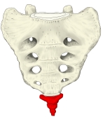 Sacrum and coccyx (anterior view)
 Vertebral column (posterior view)
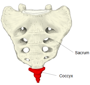
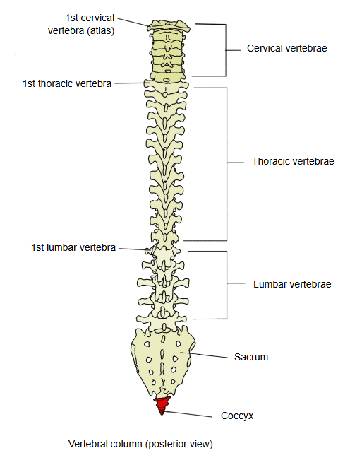
In comparative anatomy they are called caudal vertebrae:
they are the bones of the tail and articulate with the sacral vertebrae. Their number varies with the species (a rabbit, for example, has about 15), whereas in humans the few that remain are fused into one bone, the coccyx.
Where it is found
Related terms
References and sources
- Further reading: Coccyx, Wikipedia.
- Further reading: Vertebral column, Wikipedia.
- Federative International Programme for Anatomical Terminology (FIPAT). Terminologia Anatomica, 2nd ed. (TA2), 2019.