Latin: os sacrum
Overview
- Formed by the fusion of five sacral vertebrae and their rib (costal) elements; it is the largest bone of the vertebral column and forms the back wall of the pelvis.
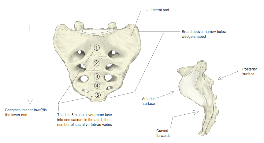
- Wedge-shaped: broad above and narrow below, thinner towards its lower end, and curved so that its pelvic surface is concave forwards.
- It has a base, an apex, a pelvic (anterior) surface, a dorsal (posterior) surface and the lateral parts.
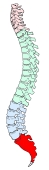
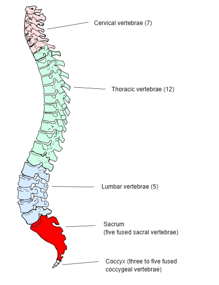
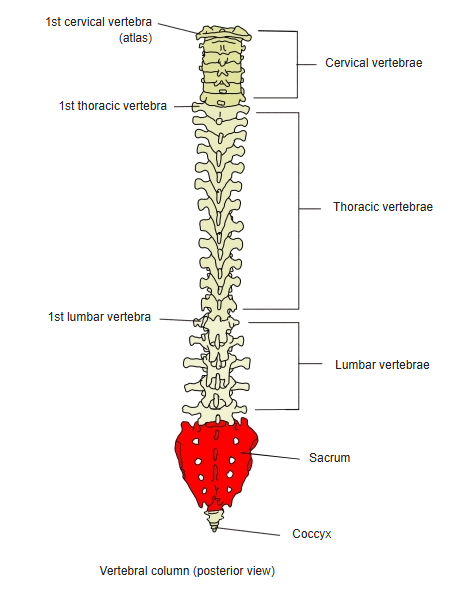
Sacrum in the vertebral column 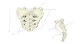
Shape of the sacrum
- Bones of the trunk
- Vertebral column
- Cervical vertebrae (7)
- Thoracic vertebrae (12)
- Lumbar vertebrae (5)
- Sacrum (1)
- Coccyx (1)
- Thoracic cage
- Sternum (1)
- Ribs (24)
- Vertebral column
Illustrations
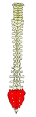
Vertebral column (posterior) 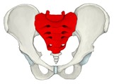
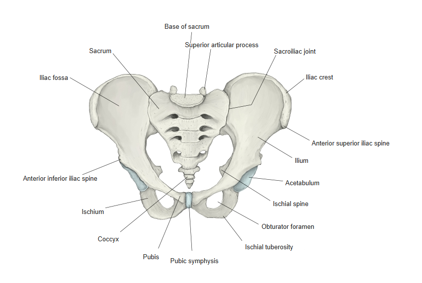
Pelvis (anterior) 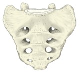
Sacrum (anterior) 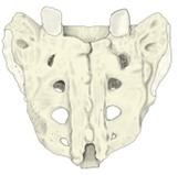
Sacrum (posterior) 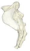
Sacrum (left lateral) 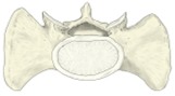
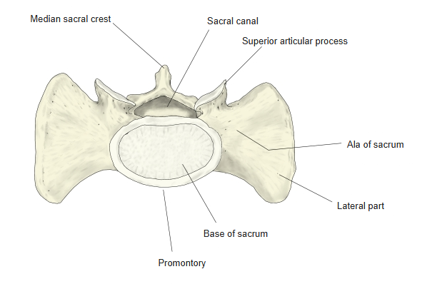
Sacrum (from above) 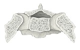
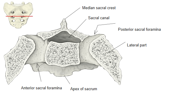
2nd sacral vertebra (horizontal section) 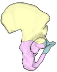
Pelvis (anterior oblique) 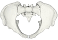
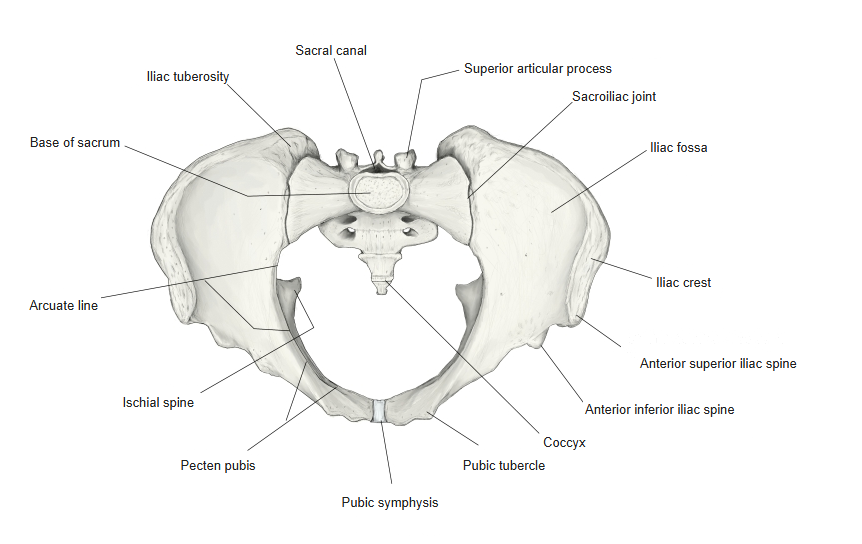
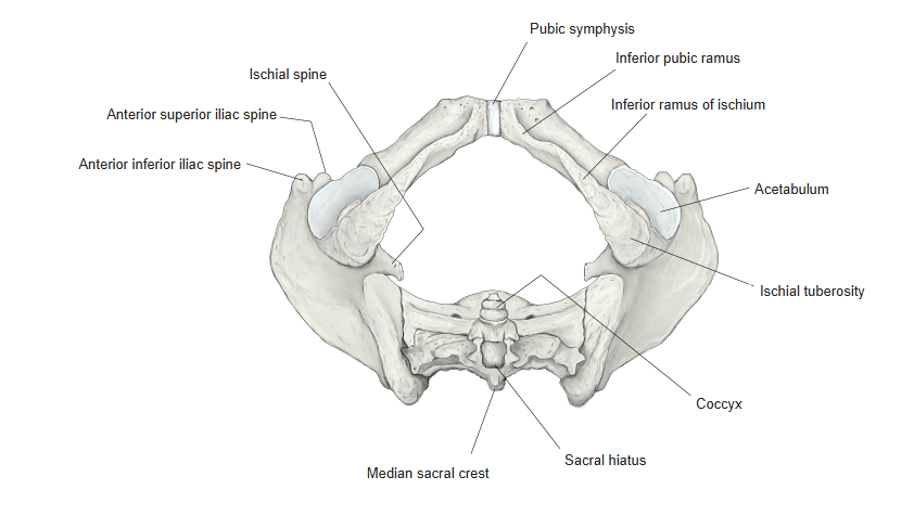
Pelvis (from above) 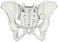
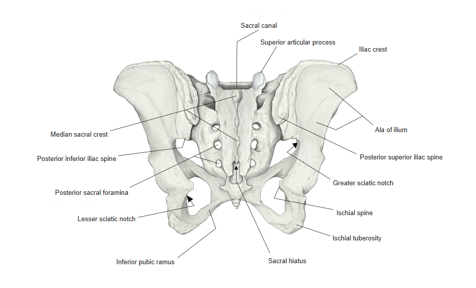
Pelvis (posterior) 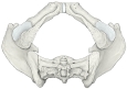
Pelvis (from below)
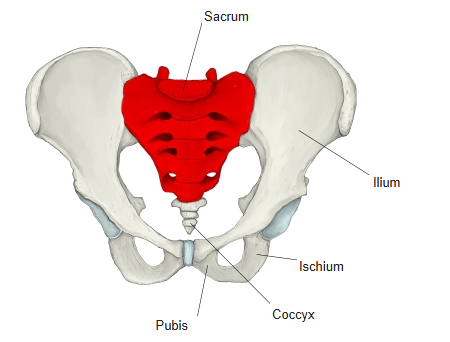
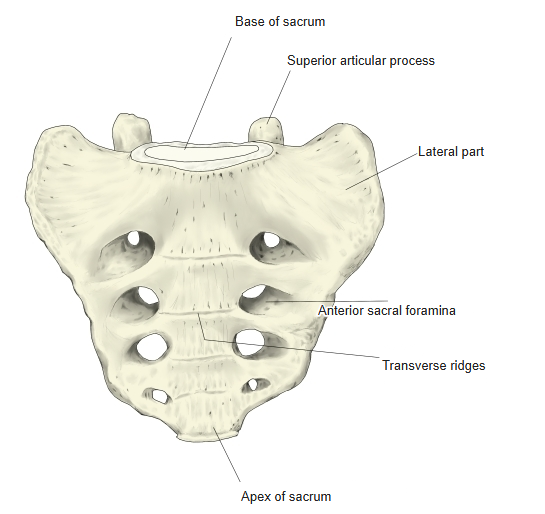
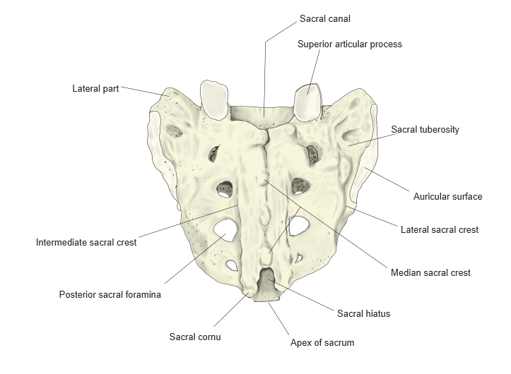
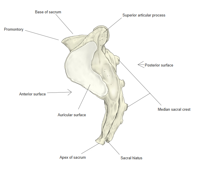
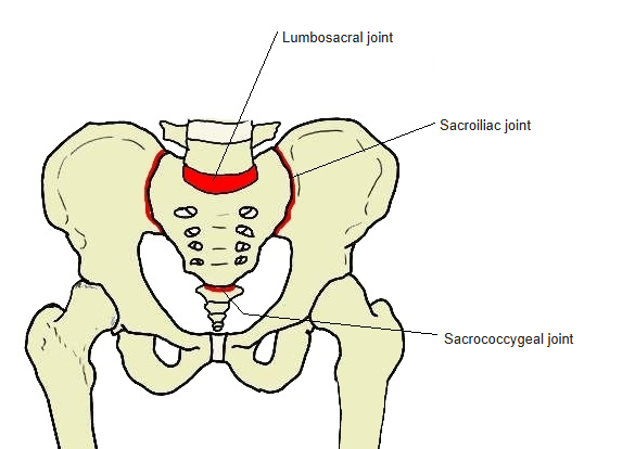
Articulations
| # | Bone | Joint |
|---|---|---|
| 1 | Ilium (auricular surface) | Sacroiliac joint |
| 2 | 5th lumbar vertebra (base of sacrum) | Lumbosacral joint |
| 3 | Coccyx (apex of sacrum) | Sacrococcygeal joint |
Joints of the sacrum
Ligaments
The following ligaments attach to the sacrum. The list is intended to be comprehensive but may omit minor ligaments.
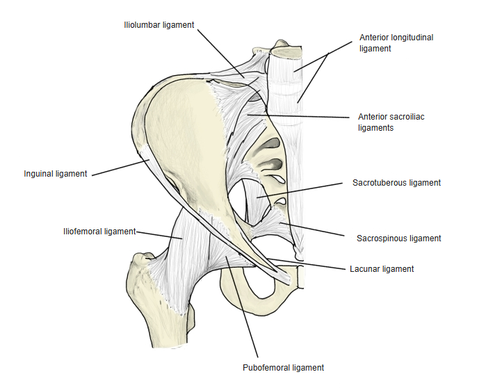
| # | Ligament | Connects |
|---|---|---|
| 1 | Anterior sacroiliac ligament | Front of the sacrum and ilium |
| 2 | Interosseous sacroiliac ligament | Between the tuberosities of the sacrum and ilium |
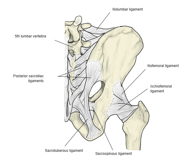
| 3 | Posterior sacroiliac ligaments (short and long) | Lateral sacral crest – posterior superior and inferior iliac spines |
| 4 | Sacrotuberous ligament | Ischial tuberosity – sacrum, coccyx and posterior inferior iliac spine |
| 5 | Sacrospinous ligament | Ischial spine – lateral margins of the sacrum and coccyx |
| 6 | Anterior longitudinal ligament | Along the front of the vertebral column from the occipital bone to the sacrum |
| 7 | Interspinous ligaments | Between the spinous processes |
| 8 | Supraspinous ligament | Tips of the spinous processes from C7 to the sacrum |
Ligaments of the pelvis (anterior) 
Ligaments of the pelvis (posterior)
Muscle attachments
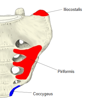
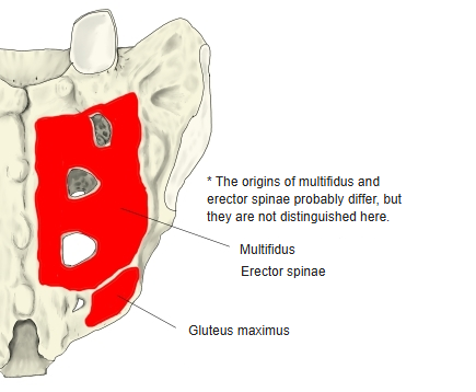
- Origin of: Piriformis, Gluteus maximus, Multifidus, Erector spinae, Iliocostalis
- Insertion of: Coccygeus
Piriformis: around the upper three anterior sacral foramina · gluteus maximus: lateral margin · multifidus and erector spinae: dorsal surface around the posterior sacral foramina · coccygeus: near the apex.
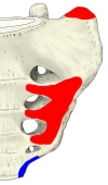
Muscle attachments (anterior) 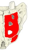
Muscle attachments (posterior) 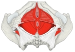
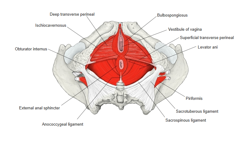
Muscles of the perineum and pelvic floor
Fusion of the sacral vertebrae
- The five sacral vertebrae are separate at first and fuse into one sacrum in the adult.
- Spinous processes → median sacral crest.
- Articular processes → intermediate sacral crests.
- Transverse processes → lateral sacral crests.
- Rib elements → lateral parts.
- Intervertebral foramina → anterior (pelvic) and posterior sacral foramina.
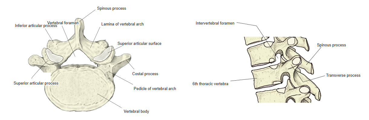
A typical vertebra
Sex differences
- The female sacrum is broader for its length, shorter and less curved, closer to an equilateral triangle; the male sacrum is longer, narrower and more strongly curved.
- The sacrococcygeal joint is movable, especially in women.
Variation
- Only about half of sacra consist of five vertebrae; six is fairly common, four is rare.
- The sacrum may be asymmetrical (for example when a sacral rib element is missing on one side or the 5th lumbar vertebra has a well-developed rib element on one side); the auricular surfaces then lie at different heights, which affects the shape of the pelvis.
References and sources
- Further reading: Sacrum, Wikipedia.