Sacrococcygeal joint (Latin: articulatio sacrococcygea)
Also called Sacrococcygeal symphysis
What is the sacrococcygeal joint?
Bones
Ligaments
The following ligaments belong to the joint, but some of them may shrink or ossify and become part of the sacrum.
| Ligament | |
|---|---|
| 1 | Anterior sacrococcygeal ligament |
| 2 | Lateral sacrococcygeal ligament |
| 3 | Superficial dorsal sacrococcygeal ligament |
| 4 | Deep dorsal sacrococcygeal ligament |
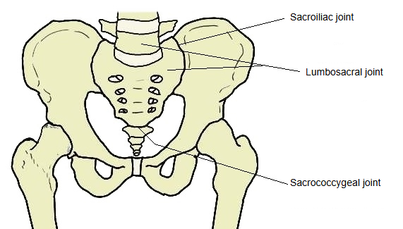
More about the joint
The ligaments of the sacrococcygeal region are reduced; some have ossified and become part of the sacrum. The main ligaments are the superficial dorsal (posterior), deep dorsal (posterior), anterior and lateral sacrococcygeal ligaments. The apex of the sacrum and the 1st coccygeal vertebra are joined by cartilage through an intervertebral disc, but this union often ossifies into a bony union. The disc sometimes contains a cavity where the nucleus pulposus would be, and especially in women a synovial-like tissue may line it.
The joint is a slightly movable symphysis between the oval surface at the apex of the sacrum and the base of the coccyx, comparable to the joints between the vertebral bodies. It is often partly or completely obliterated in old age.
- Disc: the sacrococcygeal disc is like an intervertebral disc but thinner, thicker in front and behind than at the sides, and firmer. The articular surfaces are elliptical, with their long axes running from side to side; the sacral surface is convex and the coccygeal surface concave. Sometimes the coccyx moves freely on the sacrum, particularly during pregnancy, and then a synovial membrane is present.
- Anterior sacrococcygeal ligament: a continuation of the anterior longitudinal ligament; a few irregular fibres attached to the front of the sacrum and coccyx, blending with the periosteum.
- Deep dorsal sacrococcygeal ligament: a flat band corresponding to the posterior longitudinal ligament; it runs from the back of the 5th sacral body, inside the sacral canal, to the back of the coccyx.
- Superficial dorsal sacrococcygeal ligament: corresponds to the ligamenta flava and closes the back of the lower end of the vertebral canal; it runs from the median sacral crest and the free margin of the sacral hiatus to the back of the coccyx.
- Lateral sacrococcygeal ligament: runs from the lower lateral angle of the sacrum to the transverse process of the 1st coccygeal vertebra, completing the foramen for the 5th sacral nerve.
- Intercornual ligaments: join the sacral horns to the coccygeal horns.
- Movements: limited to flexion and extension, mostly passive, during defecation and childbirth. They can add to the increase in the front-to-back diameter of the pelvic outlet produced by movement of the sacrum.
- Palpation: the joint can be felt as a horizontal groove deep in the natal (gluteal) cleft.