Coccygeal cornu (Latin: cornu coccygeum)
Also called the coccygeal horn
· It meets the sacral cornu of the sacrum, and together they leave an opening through which the fifth sacral nerve passes.
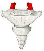 Coccyx
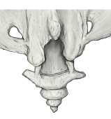 Lower sacrum and coccyx (posterior view)
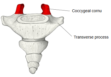
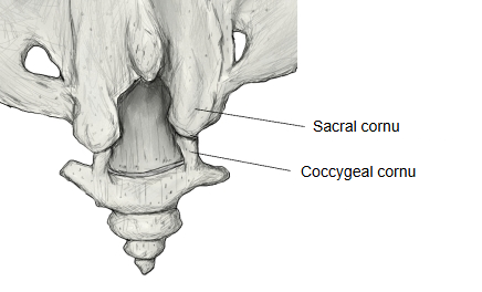
In more detail:
The two coccygeal cornua (also called coccygeal horns) project upwards from the back of the base of the coccyx. They belong to the first of the coccygeal vertebrae and are remnants of its pedicles and superior articular processes. Each cornu meets the sacral cornu of the sacrum above it, joined by ligament or by a small joint at the sacrococcygeal joint. Between the sacral and coccygeal cornua on each side is a gap through which the fifth sacral spinal nerve leaves the sacral canal. Each cornu lies just behind and medial to the small transverse process of the coccyx. Together with the sacral cornua they can be felt at the lower end of the sacral hiatus, a guide for caudal epidural injection.
Reference websites
[Wikipedia]
· Coccyx, Wikipedia.
[Websites with illustrations]
· The coccyx (TeachMeAnatomy)
· Coccyx, anterior and posterior views (Wikimedia Commons)

Where it is found
Related terms
References and sources
- Further reading: Coccyx, Wikipedia.
- Federative International Programme for Anatomical Terminology (FIPAT). Terminologia Anatomica, 2nd ed. (TA2), 2019.