Epidural space (Latin: spatium epidurale)
Also called the extradural space
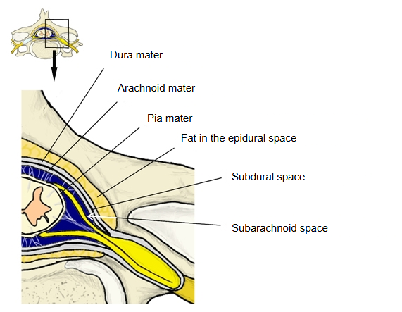
Most English-language sources use the term only for the space around the spinal dura mater:
Medical dictionaries such as The Free Dictionary define it in the same way, as the space between the walls of the vertebral canal and the dura mater of the spinal cord.
Some anatomy textbooks, however, also use the term for the skull:
In children the cranial dura mater is firmly attached to the bones of the skull. In adults it stays fused to the base of the skull and along the sutures between the skull bones, but elsewhere a very narrow space can form between the dura and the bone; this is also called the epidural space. It contains the vessels that supply the dura mater, such as the middle meningeal artery, and the sensory nerves of the dura.
In the vertebral canal the epidural space is a real space. It reaches from the foramen magnum, where the spinal dura is fused to the margin of the foramen, down to the sacral hiatus, closed by the sacrococcygeal membrane. It is filled with fat (adipose tissue) and the internal vertebral venous plexuses (anterior and posterior), and the spinal nerve roots cross it. In the head it is normally only a potential space; bleeding from a torn artery, typically the middle meningeal artery, can strip the dura from the bone and form an epidural (extradural) hematoma.
- Epidural (extradural) space : the space between the spinal dura mater and the wall of the vertebral canal
- Dura mater
- Subdural space : appears only in pathological conditions, such as after an injury
- Arachnoid mater
- Subarachnoid space : filled with cerebrospinal fluid
- Pia mater
Reference websites
[Wikipedia]
· Epidural space, Wikipedia.
· Epidural hematoma, Wikipedia.
[Websites with illustrations]
· Diagrammatic transverse section of the spinal cord and its membranes (Wikimedia Commons)

· The meninges: dura, arachnoid and pia mater (TeachMeAnatomy)