Filtration slit
Also called the slit pore
Overview
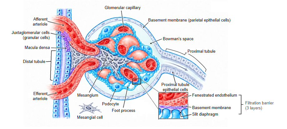
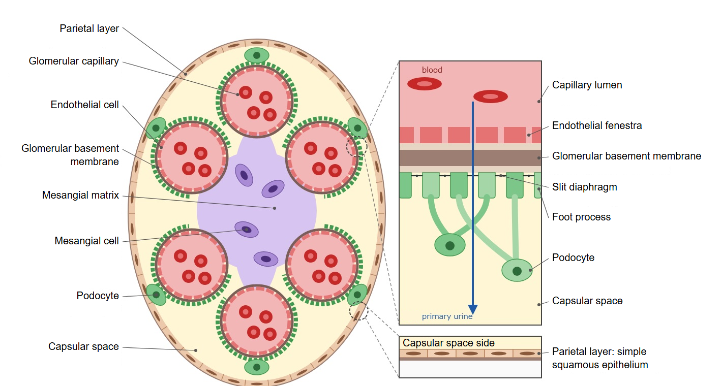
· A filtration slit is one of the narrow gaps between the foot processes of neighbouring podocytes. The slits lie in rows on the outer surface of the glomerular basement membrane. They are also called slit pores.
· The foot processes of one podocyte interlock alternately with the foot processes of the next, so the filtration slits form countless long, narrow gaps over the surface of the glomerular capillaries. Their width is usually given as about 40 nm (sources differ).
· Near the basement membrane, each filtration slit is bridged by a thin membrane, the slit diaphragm. The slit diaphragm is the membrane that closes the floor of the gap, and it is distinguished from the filtration slit, which is the gap itself.
· Blood plasma passes in turn through the pores of the endothelial cells, the glomerular basement membrane and the filtration slits (the slit diaphragms), becomes primary urine, and enters the capsular space (Bowman's space).
· When podocytes contract, the filtration slits narrow, the area available for filtration falls, and the glomerular filtration rate decreases. When the foot processes are lost (effacement) and the filtration slits disappear, protein leaks into the urine (proteinuria).
Reference websites
[Wikipedia]
· Podocyte, Wikipedia.
· Glomerulus (kidney), Wikipedia.
[Websites with illustrations]
· Filtration barrier (diagram) (Wikimedia Commons)

· Podocytes (Wikimedia Commons)

· Renal corpuscle (electron micrograph) (Histology Guide)
Related terms
- Podocyte
- Foot process
- Slit diaphragm
- Glomerular basement membrane
- Glomerular capillary
- Capsular space
- Glomerular filtration rate
References and sources
- Further reading: Podocyte, Wikipedia.
- Further reading: Glomerulus (kidney), Wikipedia.
- Federative International Programme for Anatomical Terminology (FIPAT). Terminologia Anatomica, 2nd ed. (TA2), 2019.