Slit diaphragm
Overview
· The slit diaphragm is a very thin, membrane-like structure that bridges the gap (the filtration slit) between the foot processes of neighbouring podocytes. It spans only the gaps between foot processes and lies just outside the glomerular basement membrane, near the level where the foot processes attach to it. It is not a membrane covering the foot processes.
· It is the last of the three filtering layers that blood in the glomerular capillaries passes through: the pores of the endothelial cells, the glomerular basement membrane and the slit diaphragm. Fluid that has passed the slit diaphragm enters the capsular space (Bowman's space) as primary urine.
· It is formed by proteins such as nephrin, which project from the foot processes on both sides and interlock in the middle of the gap like the teeth of a zipper. The mesh lets small molecules such as water, glucose and electrolytes through but holds back large proteins such as albumin. Proteins such as podocin and CD2AP anchor the slit diaphragm to the foot processes and stabilize it.
· Mutations in the nephrin gene cause congenital nephrotic syndrome of the Finnish type, with heavy proteinuria from birth. Mutations in the podocin gene also cause nephrotic syndrome.
· In some sources the gap (filtration slit) and the membrane across it (slit diaphragm) are used as the same term. On this site the gap is called the filtration slit and the membrane the slit diaphragm.
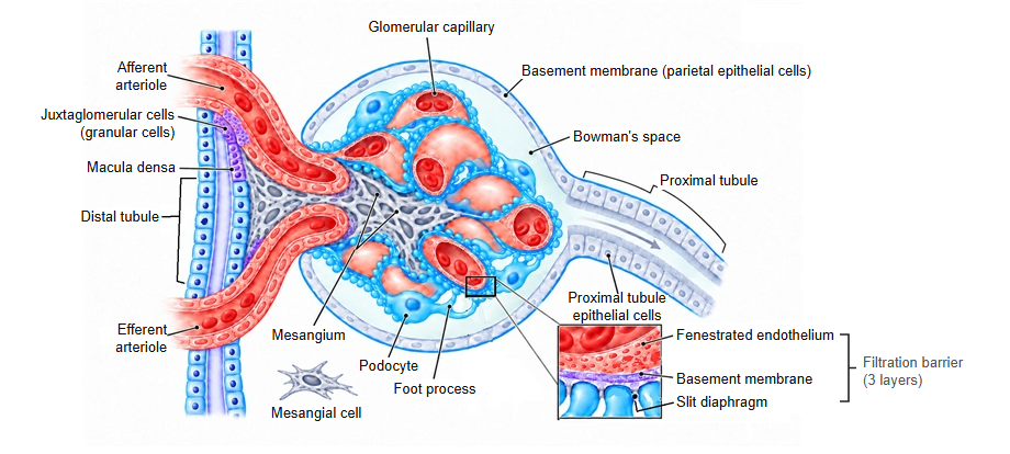
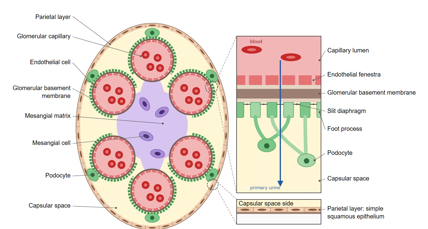
The three layers that filter the blood
Reference websites
[Wikipedia]
· Podocyte, Wikipedia.
· Nephrin, Wikipedia.
[Websites with illustrations]
· Slit diaphragm proteins of the podocyte (diagram) (Wikimedia Commons)

· Glomerular filtration barrier (diagram) (Wikimedia Commons)

· Actin cytoskeleton in the foot processes (diagram) (Wikimedia Commons)

· Renal corpuscle (electron micrograph) (Histology Guide)
Related terms
- Podocyte
- Foot process
- Filtration slit
- Glomerular basement membrane
- Endothelial cell
- Capsular space
- Glomerular capillary