Groove for middle meningeal artery (parietal bone) (Latin: sulcus arteriae meningeae mediae (os parietale))
Also called the grooves for the middle meningeal artery on the parietal bone
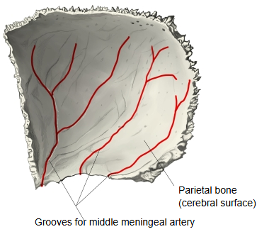
Among the arterial grooves on the cerebral surface of the parietal bone, the grooves made by the middle meningeal artery are the most prominent.
The frontal (anterior) branch reaches the bone near the sphenoidal angle, where its groove may be deep or even a short bony canal, and runs upwards and backwards; the parietal (posterior) branch grooves the lower, posterior part of the bone. Each groove divides like the branches of a tree and also holds the accompanying middle meningeal veins. Because the vessels lie in the outer layer of the dura mater close against the bone, a fracture near the sphenoidal angle (the pterion region) can tear them and cause an extradural haemorrhage. The temporal bone has a groove of the same name (groove for middle meningeal artery of the temporal bone).
Reference websites
[Wikipedia]
· Middle meningeal artery, Wikipedia.
· Parietal bone, Wikipedia.
[Websites with illustrations]
· Left parietal bone, inner surface (Gray's Anatomy, fig. 133) (Wikimedia Commons)

· Base of the skull, inner surface (Gray's Anatomy, fig. 193) (Wikimedia Commons)

Related terms
- Arterial grooves
- Parietal bone
- Middle meningeal artery
- Sphenoidal angle of parietal bone
- Dura mater
- Groove for superior sagittal sinus
- Groove for middle meningeal artery (temporal bone)
References and sources
- Further reading: Middle meningeal artery, Wikipedia.
- Federative International Programme for Anatomical Terminology (FIPAT). Terminologia Anatomica, 2nd ed. (TA2), 2019.