Groove for middle meningeal artery (temporal bone) (Latin: sulcus arteriae meningeae mediae)
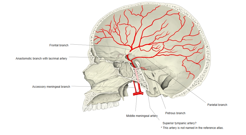
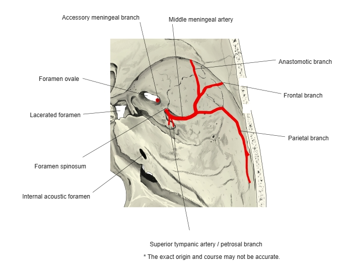
· The parietal bone has a groove of the same name. The middle meningeal artery divides on the temporal squama into a frontal (anterior) and a parietal (posterior) branch, and both branches leave clear grooves that run on from the temporal bone onto the greater wing of the sphenoid and the parietal bone. Because the bone here is thin, a fracture can tear the artery and cause an extradural (epidural) haemorrhage.
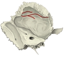 Left temporal bone (medial surface)
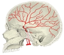 Middle meningeal artery (1)
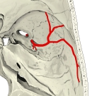 Middle meningeal artery (2)
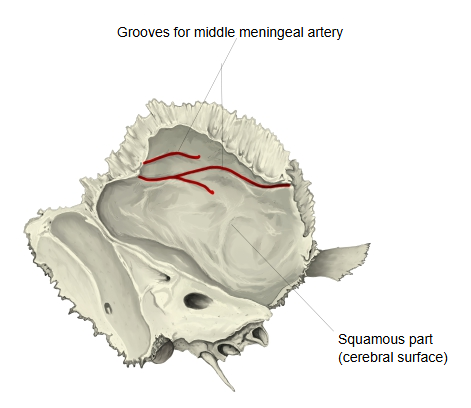
Reference websites
[Wikipedia]
· Middle meningeal artery, Wikipedia.
· Temporal bone, Wikipedia.
[Websites with illustrations]
· Left temporal bone, inner surface (Gray's Anatomy, fig. 138) (Wikimedia Commons)

· Base of the skull, inner surface (Gray's Anatomy, fig. 193) (Wikimedia Commons)

Related terms
- Arterial grooves
- Squamous part
- Groove for middle temporal artery
- Foramen spinosum
- Middle cranial fossa
- Dura mater
- Temporal bone
- Groove for middle meningeal artery (parietal bone)
References and sources
- Further reading: Middle meningeal artery, Wikipedia.
- Further reading: Temporal bone, Wikipedia.
- Federative International Programme for Anatomical Terminology (FIPAT). Terminologia Anatomica, 2nd ed. (TA2), 2019.