Inferior vertebral notch (Latin: incisura vertebralis inferior)
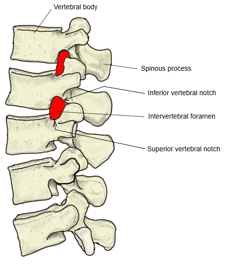
· With the superior vertebral notch of the vertebra just below, it forms the intervertebral foramen, through which the spinal nerve passes.
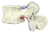 Lumbar vertebra (lateral view)
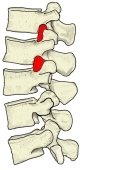 Lumbar vertebrae and intervertebral foramina
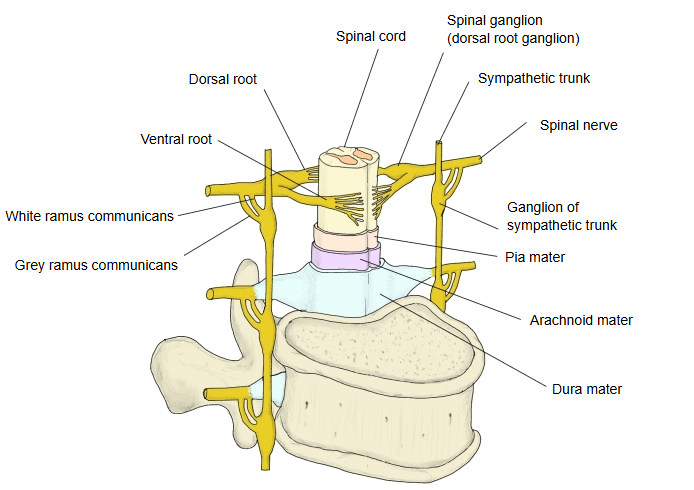
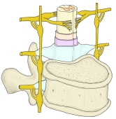 Spinal cord, meninges and spinal nerves
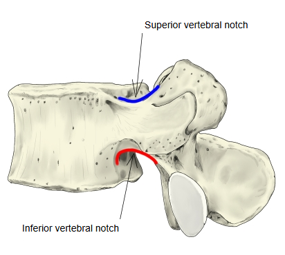
In more detail:
Each pedicle joins the vertebral body to the vertebral arch. Its upper border has a shallow superior vertebral notch and its lower border a deeper inferior vertebral notch. In the thoracic and lumbar vertebrae the inferior notch is much deeper than the superior one, so it forms most of the intervertebral foramen; in the cervical vertebrae the two notches are more equal. The foramen is completed in front by the intervertebral disc and behind by the zygapophysial joint. The spinal nerve, its spinal (dorsal root) ganglion and small vessels pass through it. Narrowing of the foramen, for example by a bulging disc or bony overgrowth, can press on the spinal nerve.
Reference websites
[Wikipedia]
· Intervertebral foramen, Wikipedia.
· Vertebra, Wikipedia.
[Websites with illustrations]
· The vertebral column (TeachMeAnatomy)
· A lumbar vertebra from above and behind (Gray's Anatomy) (Wikimedia Commons)

Related terms
- Vertebral body
- Vertebral column
- Inferior articular facet of vertebra
- Lumbar vertebrae
- Thoracic vertebrae
- Cervical vertebrae
- Spinal nerves
References and sources
- Further reading: Intervertebral foramen, Wikipedia.
- Further reading: Vertebra, Wikipedia.
- Federative International Programme for Anatomical Terminology (FIPAT). Terminologia Anatomica, 2nd ed. (TA2), 2019.