Latin: vertebrae lumbales · L1–L5
Overview
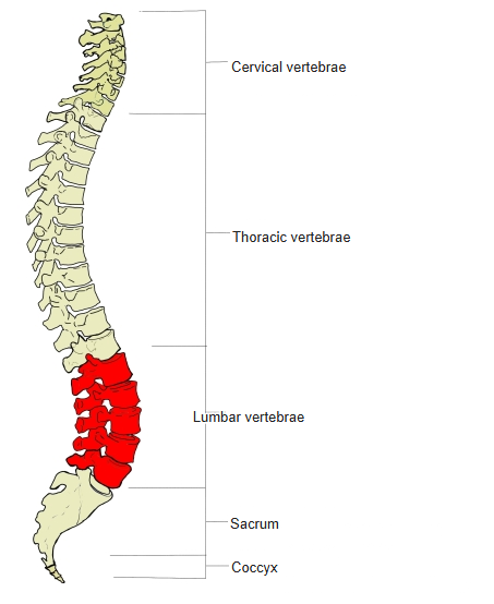
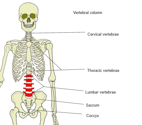
- The five lumbar vertebrae lie below the thoracic vertebrae; they are numbered from above, L1 to L5.
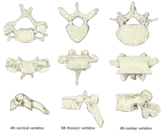
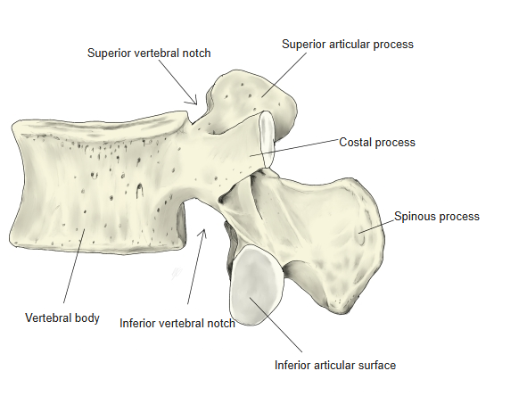
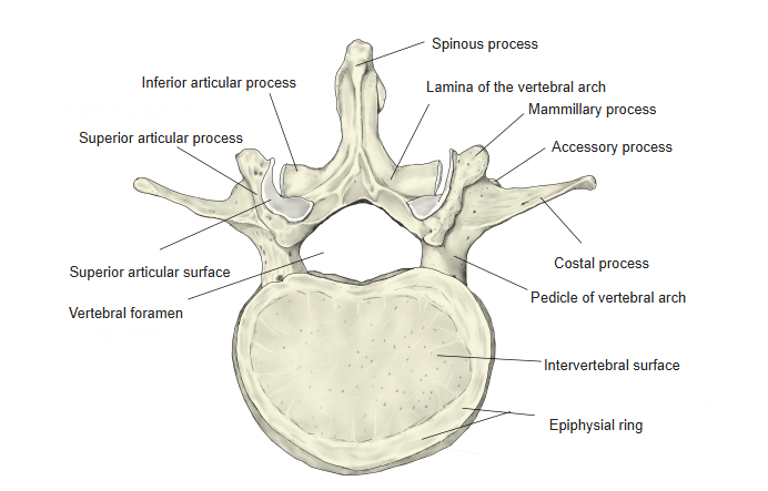
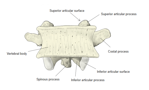
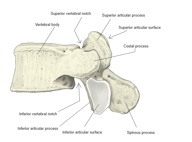
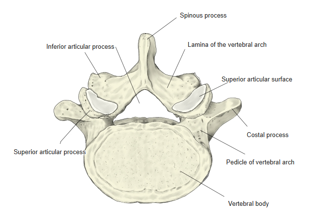
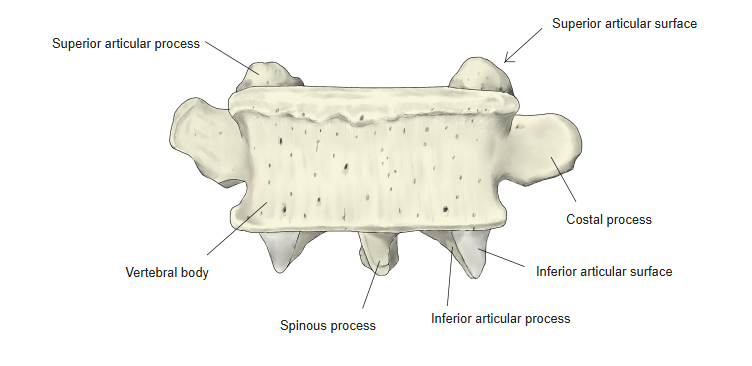
- The flat process that projects laterally from the pedicle, in the position of a transverse process, is the costal process; it corresponds to a rib in this region and is larger in the lower lumbar vertebrae.
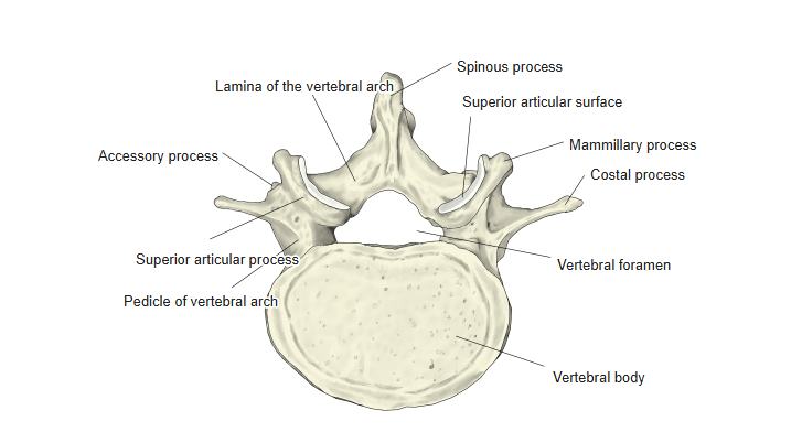
- The true transverse process remains as two small elevations: the mamillary process (on the posterior border of the superior articular process) and the accessory process (a small spine on the back of the root of the costal process, which gives attachment to muscles).
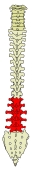
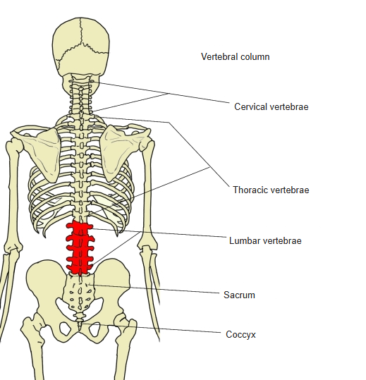
Lumbar vertebrae in the vertebral column (posterior) 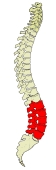
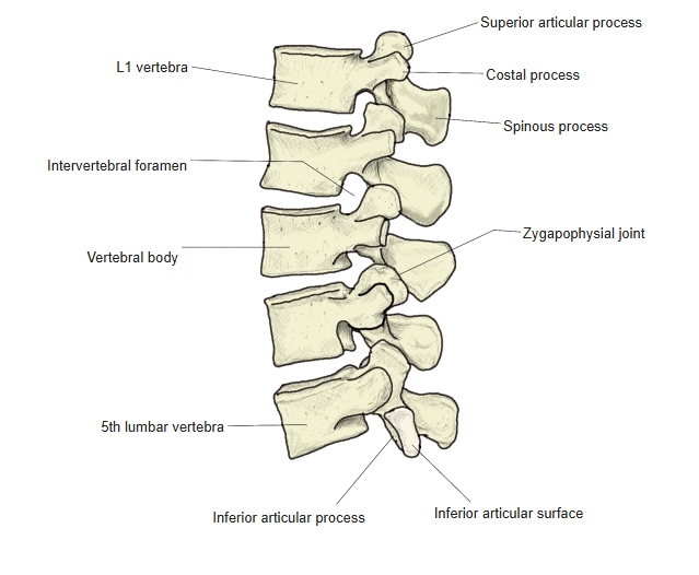
Lumbar vertebrae in the vertebral column (lateral) 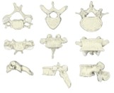
Cervical, thoracic and lumbar vertebrae compared
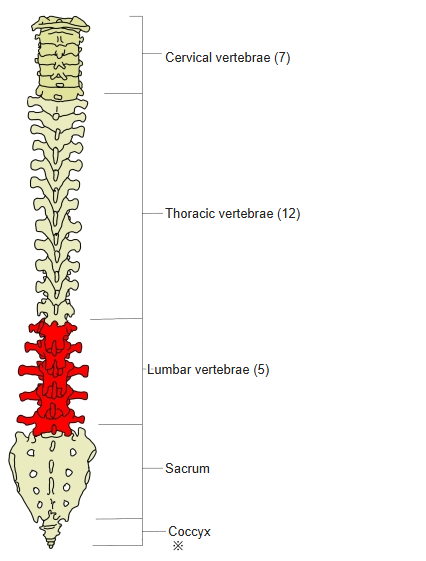
- Bones of the trunk
- Vertebral column
- Cervical vertebrae (7)
- Thoracic vertebrae (12)
- Lumbar vertebrae (5)
- Sacrum (1)
- Coccyx (1)
- Thoracic cage
- Sternum (1)
- Ribs (24)
- Vertebral column
Illustrations
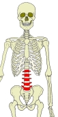
Position of the lumbar vertebrae (anterior) 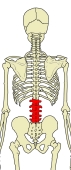
Position of the lumbar vertebrae (posterior) 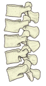
Lumbar vertebrae (left lateral) 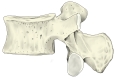
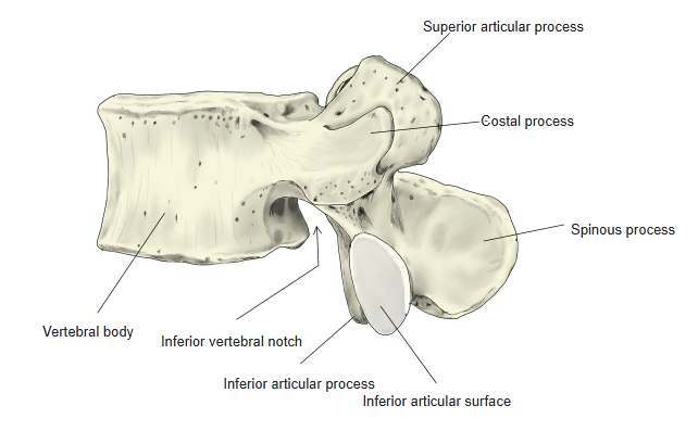
2nd lumbar vertebra (left lateral) 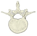
2nd lumbar vertebra (from above) 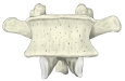
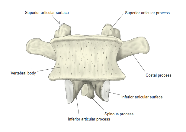
2nd lumbar vertebra (anterior) 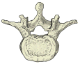
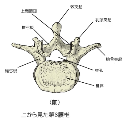
3rd lumbar vertebra (from above) 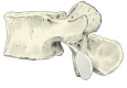
4th lumbar vertebra (left lateral) 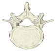
4th lumbar vertebra (from above) 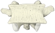
4th lumbar vertebra (anterior) 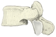
5th lumbar vertebra (left lateral) 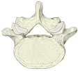
5th lumbar vertebra (from above) 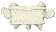
5th lumbar vertebra (anterior)
Articulations
| # | Bone | Joint |
|---|---|---|
| 1 | Inferior articular surface ⇔ superior articular surface of the next vertebra | Zygapophysial (facet) joints |
| 2 | Vertebral bodies (through the intervertebral discs) | Intervertebral joints (symphyses) |
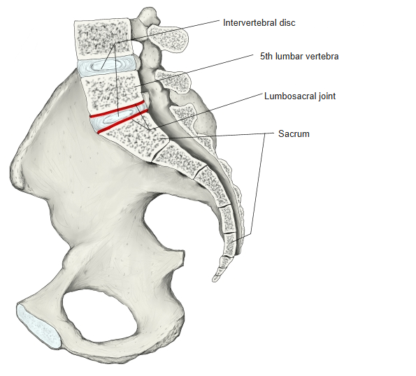
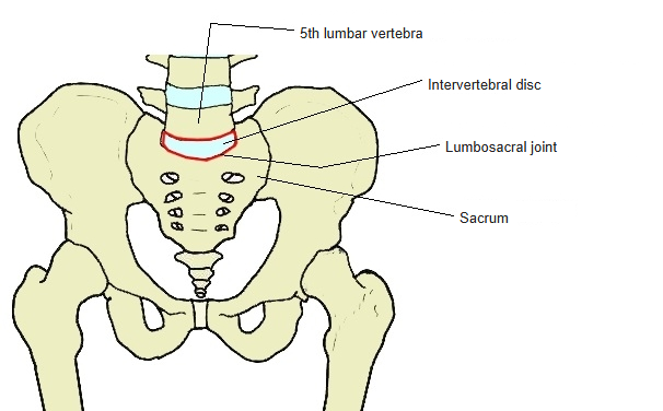
| 3 | 5th lumbar vertebra ⇔ sacrum | Lumbosacral joint |
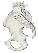
Lumbosacral joint (median section) 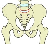
Lumbosacral joint (anterior)
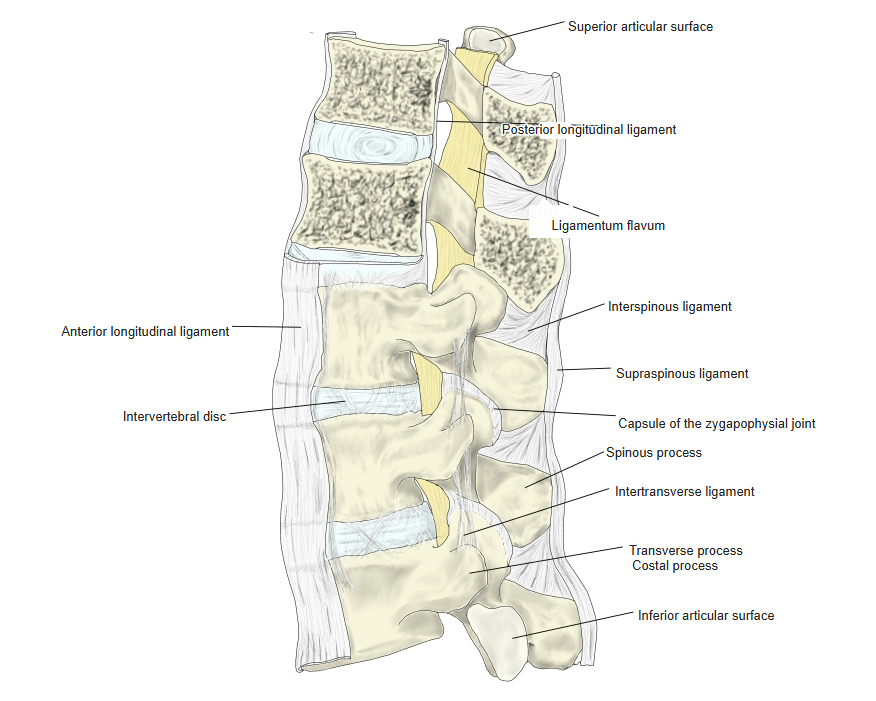
Ligaments
The following ligaments attach to the lumbar vertebrae. The list is intended to be comprehensive but may omit minor ligaments.
| # | Ligament | Connects |
|---|---|---|
| 1 | Anterior longitudinal ligament | Occipital bone (pharyngeal tubercle) – front of the sacrum |
| 2 | Intertransverse ligaments | Between the transverse (costal) processes |
| 3 | Posterior longitudinal ligament | Occipital bone (clivus) – sacral canal |
| 4 | Ligamenta flava | Between the laminae |
| 5 | Interspinous ligaments | Between the spinous processes |
| 6 | Supraspinous ligament | Tips of the spinous processes |
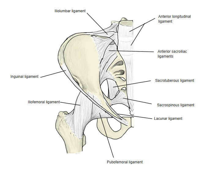
| 7 | Iliolumbar ligament | Costal processes of L4 and L5 – iliac crest |
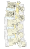
Ligaments of the lumbar spine 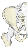
Ligaments of the pelvis (anterior)
Muscle attachments
- Origin of: Latissimus dorsi, Interspinales, Iliocostalis, Longissimus, Intertransversarii, Spinalis, Multifidus, Psoas minor, Rotatores, Psoas major
- Insertion of: Quadratus lumborum
Multifidus: mamillary and accessory processes · psoas major: vertebral bodies and costal processes · quadratus lumborum: costal processes. Muscles that both arise from and insert on lumbar vertebrae are listed under origin.
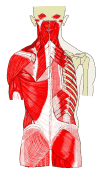
Muscles of the back (superficial and deep) 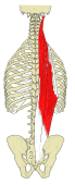
Erector spinae 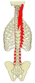
Splenius and intertransverse muscles 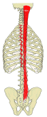
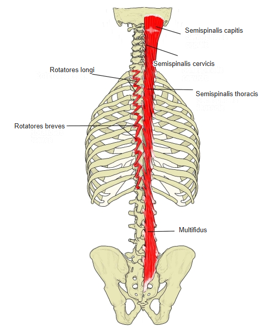
Transversospinales 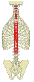
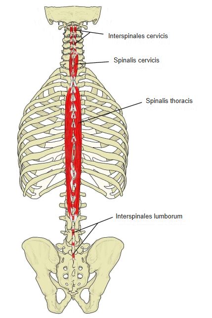
Spinalis and interspinales
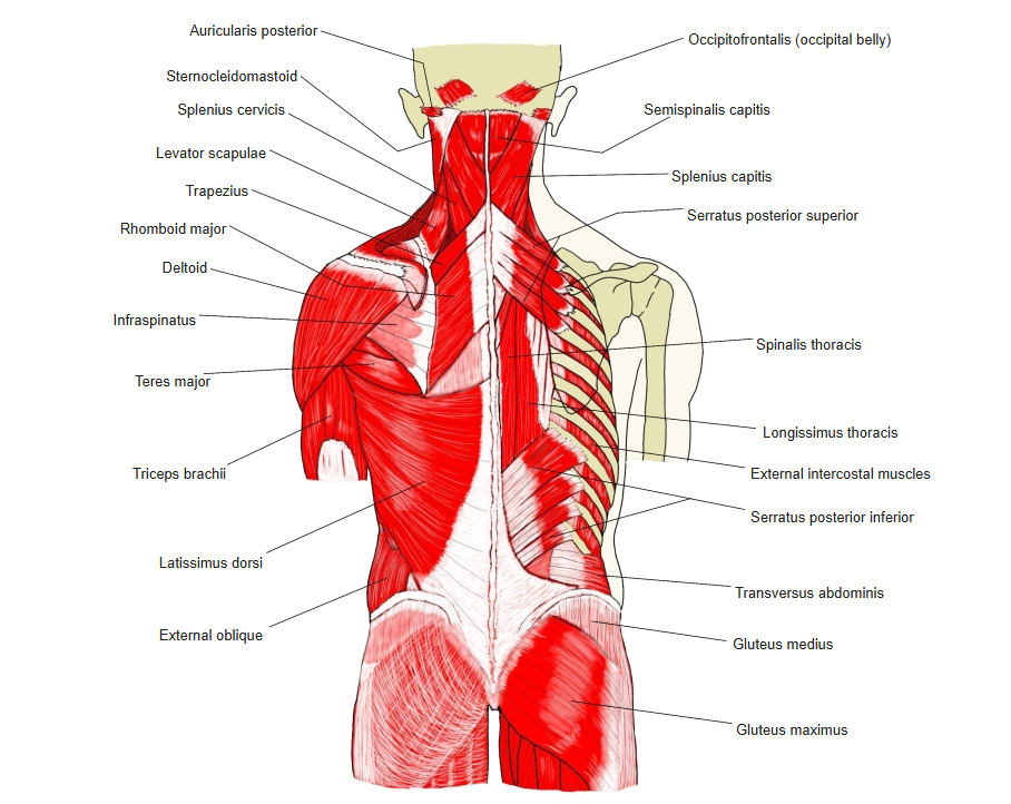
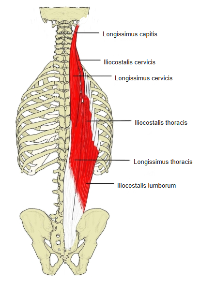
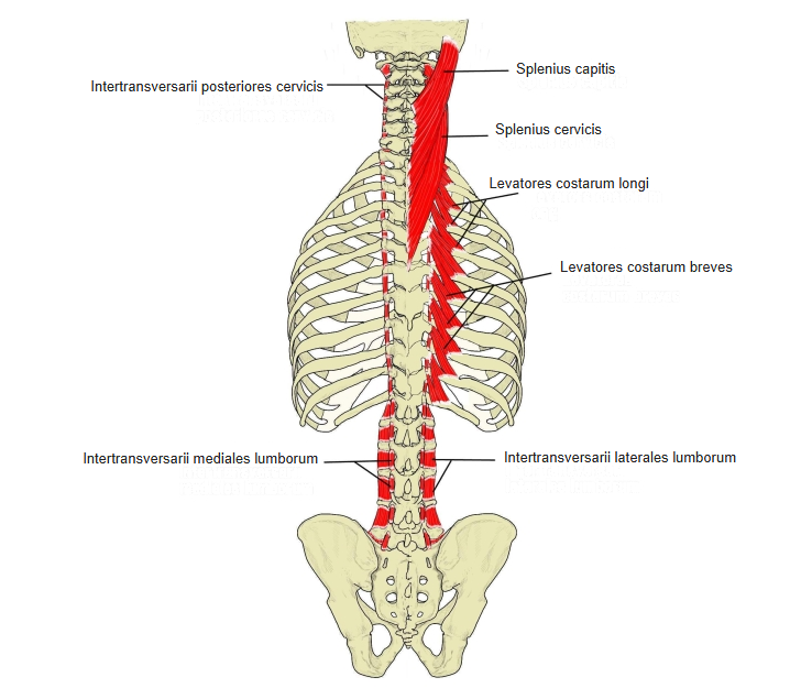
Parts of a lumbar vertebra
- Vertebral body – the largest of all the vertebrae; wider in the lower ones, and thickest at about L3 or L4.
- Costal process – takes the place of the transverse process of other vertebrae; the true transverse process survives as the mamillary and accessory processes.
- Spinous process – short, broad and box-shaped, projecting almost horizontally backwards.
- Superior and inferior articular processes – project vertically upwards and downwards.
- Vertebral foramen – triangular, and larger than in the thoracic vertebrae.
References and sources
- Further reading: Lumbar vertebrae, Wikipedia.