Renal parenchyma (Latin: parenchyma renis)
Overview
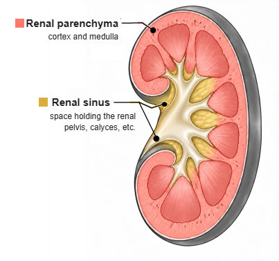
· The parenchyma surrounds the renal sinus, the central space that opens at the renal hilum. Its outer surface is covered by the fibrous capsule, and on the inside the renal papillae project into the minor calyces.
· The calyces, the renal pelvis, and the vessels, nerves and fat inside the sinus are not part of the parenchyma.
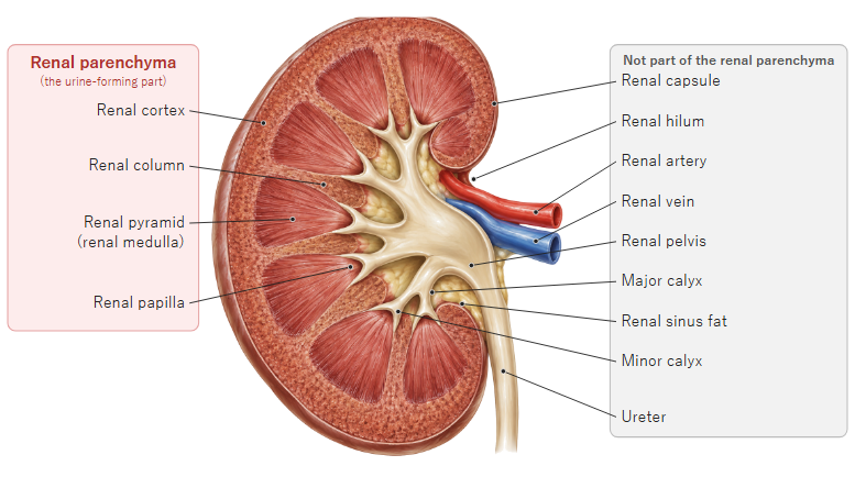
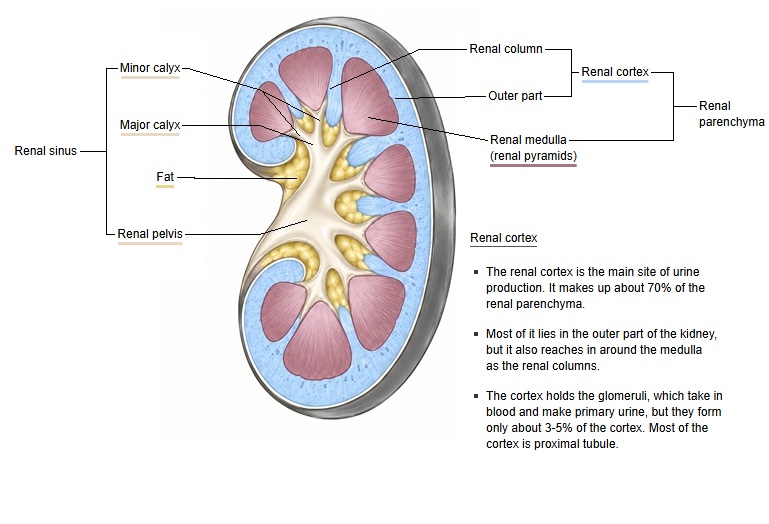
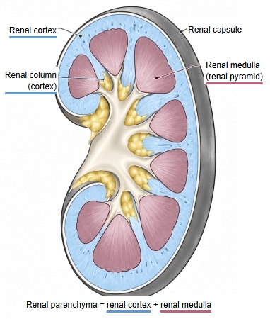
- Renal parenchyma : the part that forms urine (cortex and medulla).
- Renal cortex : the outer layer just beneath the capsule; renal corpuscles and convoluted tubules gather here.
- Renal column : cortex that extends between the renal pyramids.
- Renal medulla : inside the cortex; made of several (roughly 8-18) renal pyramids.
- Renal pyramid : the cone-shaped part where the loops of Henle and collecting ducts gather.
- Renal papilla : the tip of the pyramid; papillary ducts open here and pass urine into a minor calyx.
- Renal cortex : the outer layer just beneath the capsule; renal corpuscles and convoluted tubules gather here.
- Other parts : everything outside the renal parenchyma.
- Renal sinus : the inner space continuing from the hilum; it contains:
- Calyces and pelvis : minor calyx → major calyx → renal pelvis → to the ureter.
- Vessels and nerves : branches of the renal artery and vein, the renal nerve plexus.
- Fat : fills the gaps between the vessels and calyces.
- Renal hilum : the notch on the medial border; the renal artery, renal vein and pelvis pass through it.
- Coverings : the layers around the kidney, from inside out: fibrous capsule → perirenal fat capsule → renal fascia.
- Renal sinus : the inner space continuing from the hilum; it contains:
· The medulla is made up of the renal pyramids (roughly 8 to 18; sources differ). Cortex dips between the pyramids as the renal columns. One pyramid with the cortex over it forms a renal lobe.
· The units that make urine, the nephrons and collecting ducts, lie within the parenchyma: the renal corpuscles are in the cortex, and the loops of Henle and most of the collecting ducts run in the medulla.
· In chronic kidney disease the parenchyma becomes thinner and the kidney shrinks. Diseases centred on this tissue are called renal parenchymal diseases.
- Renal cortex
- Renal corpuscle : glomerulus (a tuft of glomerular capillaries) and Bowman's capsule
- Proximal tubule : reabsorbs water and nutrients from the primary urine (convoluted part)
- Distal tubule : adjusts salts and water (convoluted part)
- Cortical collecting duct : collects the primary urine from several connecting tubules
- Renal medulla
- Renal pyramid : the cone-shaped tissue that forms the medulla; it contains:
- Loop of Henle : U-shaped part (proximal straight tubule, intermediate tubule, distal straight tubule)
- Medullary collecting duct : carries the primary urine toward the renal papilla
- Papillary duct : opens at the tip of the renal papilla and empties the primary urine into a minor calyx, where it becomes urine
- Renal pyramid : the cone-shaped tissue that forms the medulla; it contains:
Reference websites
[Wikipedia]
· Kidney, Wikipedia.
· Renal cortex, Wikipedia.
· Renal medulla, Wikipedia.
[Websites with illustrations]
· The anatomy of the kidney (Interactive Biology)
· Gross anatomy of the kidney (Lumen Learning, Anatomy and Physiology II)
· Kidney cross-section (EBM Consult)
· Kidney anatomy (Blausen) (Wikimedia Commons)

Related terms
- Cortical labyrinth
- Medullary rays
- Renal sinus
- Kidney
- Renal pelvis
- Fibrous capsule of kidney
- Capsule
- Perirenal fat capsule
- Renal fascia
- Ureter
References and sources
- Further reading: Kidney, Wikipedia.
- Further reading: Renal cortex, Wikipedia.
- Further reading: Renal medulla, Wikipedia.
- Federative International Programme for Anatomical Terminology (FIPAT). Terminologia Anatomica, 2nd ed. (TA2), 2019.