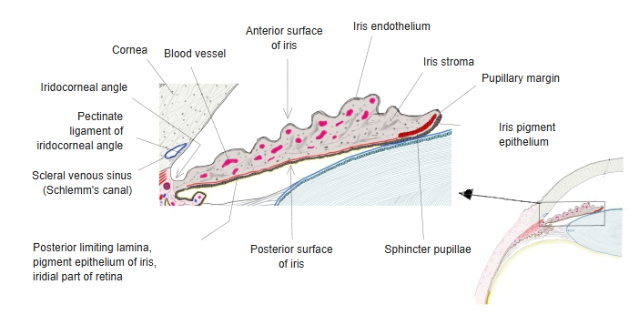
Iris endothelium
Also called the endothelium of anterior chamber
of the
iris
- Iris endothelium : = the endothelium of the anterior chamber; described as a layer of flattened polygonal cells continuing the endothelium on the back of the cornea; in the adult human iris it is not a continuous layer
- Stroma of iris
- Anterior border layer : fairly dense connective tissue with pigment cells
- Vascular layer : the main part of the iris, rich in vessels and nerves; it contains the sphincter pupillae (the dilator pupillae lies at its back)
- Posterior limiting lamina
- Behind it : the pigmented epithelium of the iris, which belongs to the inner layer of the eyeball (the iridial part of the retina)
The table above follows one classic description. Wikipedia, as below, divides the iris broadly into two layers, the stroma and the pigmented epithelial cells, and has no layer corresponding to the iris endothelium; other sources on the sense organs do not use the names iris endothelium or endothelium of the anterior chamber either.
In that description the iris has a pigmented, fibrous and vascular front layer, the stroma, and behind it a layer of pigmented epithelial cells. In the adult human eye no continuous endothelium is found on the front of the iris; the name is an older one, for the flat cells once thought to continue the endothelium of the cornea across the iridocorneal angle onto the iris.