Vascular layer of iris
of the
iris
- Iris endothelium : described as a layer of flattened cells continuing the corneal endothelium (the endothelium of the anterior chamber); in the adult human iris it is not a continuous layer
- Stroma of iris
- Anterior limiting lamina : fairly dense connective tissue with pigment cells
- Vascular layer : the main part of the iris, rich in vessels and nerves; it contains the sphincter pupillae (the dilator pupillae lies at its back)
- Posterior limiting lamina
- Behind it : the pigmented epithelium of the iris, which belongs to the inner layer of the eyeball (the iridial part of the retina)
Its connective tissue fibres (collagen fibres with a very few elastic fibres) gather mainly around the vessels (arteries and veins) and nerves, forming strong sleeves (an adventitial layer) around them. Circular fibres are rare; most of the fibres run in the same direction as the vessels.
Vessels: the major and minor arterial circles of the iris and the small arteries that link them; they carry blood to the whole iris.
Nerves: the sphincter pupillae is supplied by parasympathetic fibres of the oculomotor nerve, which reach it through the short ciliary nerves;
the dilator pupillae is supplied by sympathetic fibres that reach the eye through the long ciliary nerves and short ciliary nerves.
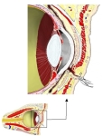 Front of the orbit (section)
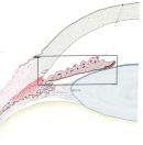 Iris (section)
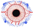 Vessels of the iris
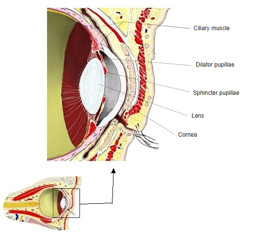
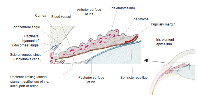
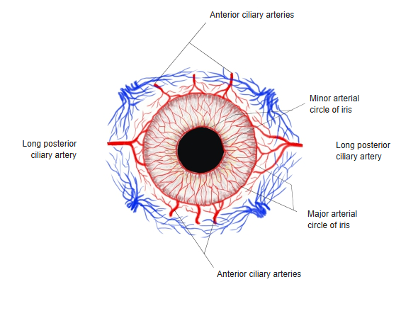
The vascular layer forms most of the iris. It is made of loose connective tissue containing pigment cells and is rich in blood vessels and nerves; it is part of the vascular layer of the eyeball (uvea). In its back part lies the sphincter pupillae, a ring of smooth muscle along the pupillary margin that narrows the pupil when it contracts (parasympathetic supply). Behind the layer, the dilator pupillae spreads as a thin sheet along the front of the pigment epithelium and runs radially from the ciliary margin to the pupillary margin; it widens the pupil when it contracts (sympathetic supply). Both muscles develop from ectoderm. The cells of the dilator belong to the pigmented epithelium of the iris: they keep epithelial features and contain pigment granules. Behind the vascular layer lie the posterior limiting lamina and then the pigmented epithelium of the iris, the iridial part of the retina, which belongs to the inner layer of the eyeball.
Related terms
- Blood
- Iris
- Anterior limiting lamina of iris
- Posterior limiting lamina of iris
- Major arterial circle of iris
- Minor arterial circle of iris
- Pupil
- Vascular layer of eyeball
References and sources
- Further reading: Iris (anatomy), Wikipedia.
- Federative International Programme for Anatomical Terminology (FIPAT). Terminologia Anatomica, 2nd ed. (TA2), 2019.