Macula of saccule (Latin: macula sacculi)
Also called the saccular macula
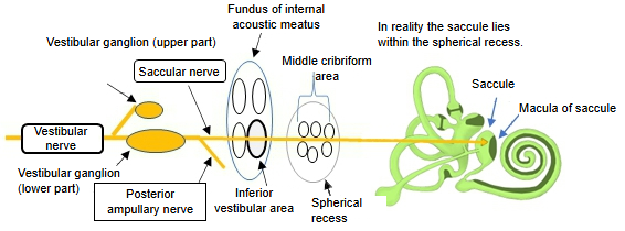
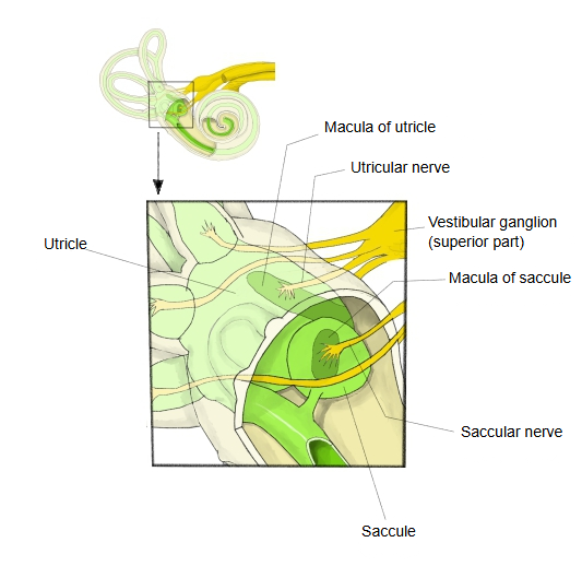
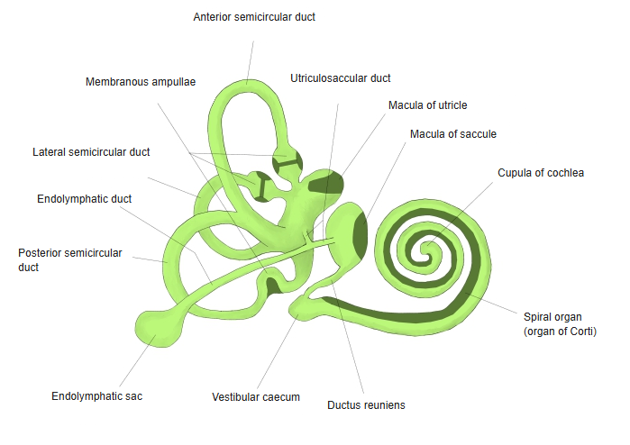
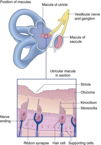
· It lies in the front part of the saccule and stands almost vertically, at right angles to the roughly horizontal macula of the utricle (utricle). Together the two are the vestibular maculae.
· Its epithelium is made of hair cells and supporting cells. The saccule itself sits in the spherical recess of the vestibule.
Hair cells of the macula of saccule
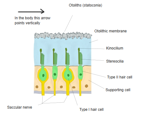
· There are two kinds of hair cells, type I and type II.
· Each hair cell carries 40 to 70 stereocilia and one kinocilium.
· The hair bundles on top of the hair cells are covered by a jelly-like otolithic membrane.
· On the surface of the otolithic membrane lie great numbers of small protein–calcium carbonate crystals (otoconia, otoliths or statoconia).
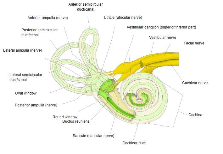
· In up-and-down movement the heavier otolithic membrane lags behind and bends the hair bundles; the hair cells pass this on to the saccular nerve, part of the vestibular nerve.
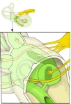 Maculae of the utricle and saccule
 Membranous labyrinth
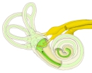 Bony and membranous labyrinth with nerves
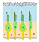 Hair cells of the macula
Reference websites
[Wikipedia]
· Macula of saccule, Wikipedia.
· Otolithic membrane, Wikipedia.
[Websites with illustrations]
· How the maculae respond to changes in head position (pharmacy180.com)

· Position of the maculae and a macula in section (Clinical Gate)
· Maculae of the utricle and saccule, with otoliths on the hair cells (nasafordoctors.co.za)

Related terms
References and sources
- Further reading: Macula of saccule, Wikipedia.
- Further reading: Otolithic membrane, Wikipedia.
- Federative International Programme for Anatomical Terminology (FIPAT). Terminologia Anatomica, 2nd ed. (TA2), 2019.