Vestibular maculae (Latin: maculae)
Also called Macula of utricle and macula of saccule
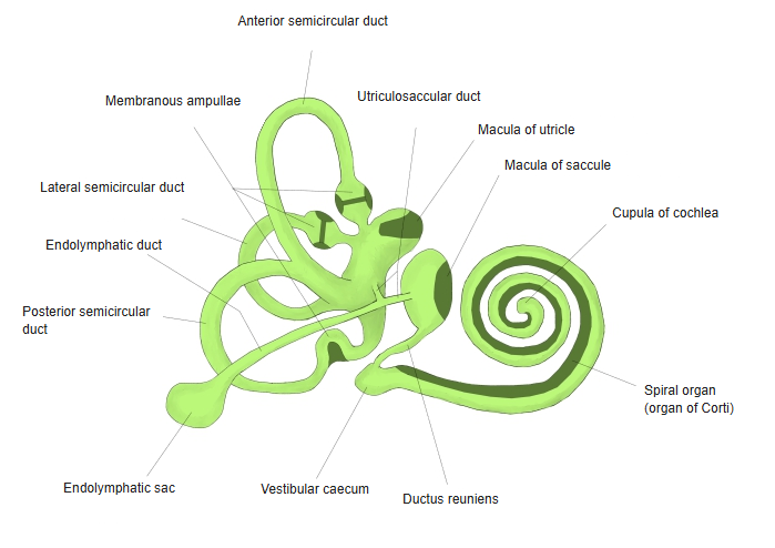
The vestibular maculae is the collective name for the macula of the saccule and the macula of the utricle, found in the two sacs of the membranous labyrinth, part of the internal ear.
The two sacs: the saccule and the utricleInternal ear: the bony labyrinth + the membranous labyrinth (cochlear duct + the two sacs + semicircular ducts)Maculae: they contain sensory hair cells
The two maculae lie inside the bony vestibule. The macula of the utricle lies roughly horizontal on the floor of the utricle; the macula of the saccule lies roughly vertical on its medial wall, so the two respond to movement in different planes.
Each is made of sensory hair cells and supporting cells, covered by a jelly-like otolithic membrane carrying tiny calcium carbonate crystals (otoconia). Gravity and straight-line acceleration shift the otolithic membrane and bend the hair bundles; the signals travel in the vestibular nerve.
Together with the ampullary crests of the semicircular ducts, which sense rotation, they form the sense organs of balance.
 Membranous labyrinth
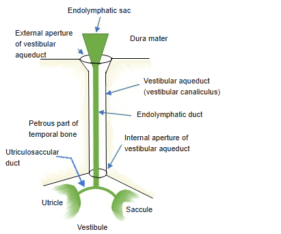
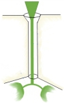 Endolymphatic duct (schematic)
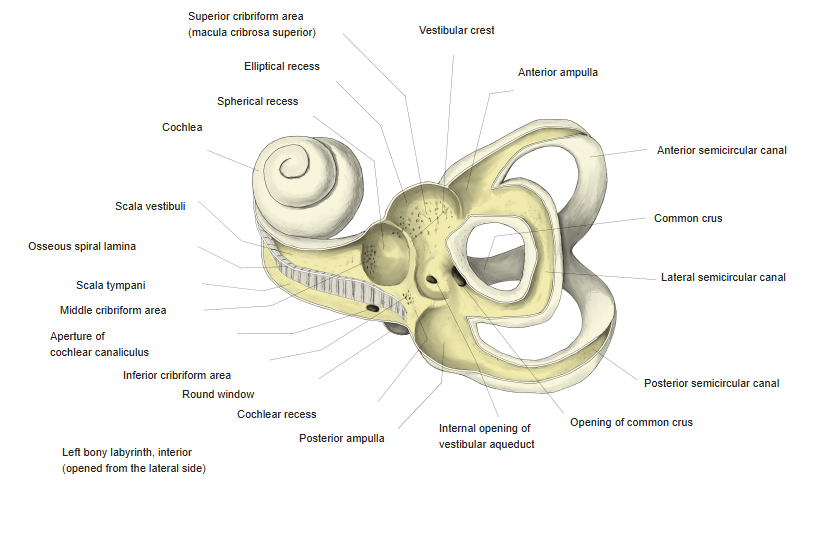
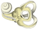 Left bony labyrinth (opened from the lateral side)
Related terms
References and sources
- Further reading: Macula of utricle, Wikipedia.
- Further reading: Otolithic membrane, Wikipedia.
- Federative International Programme for Anatomical Terminology (FIPAT). Terminologia Anatomica, 2nd ed. (TA2), 2019.