Vestibular ganglion (Latin: ganglion vestibulare)
Also called Scarpa's ganglion
Its nerve cells are bipolar. Their peripheral processes reach the hair cells of the maculae of the utricle and saccule and of the ampullary crests of the semicircular ducts; their central processes form the vestibular nerve, which runs to the vestibular nuclei in the brainstem. The superior and inferior parts are joined by a narrow isthmus.
Superior part of the vestibular ganglion
It gives the utriculoampullary nerve, which enters the vestibule through the superior vestibular area and divides into three branches:
1) Utricular nerve: to the sensory epithelium of the macula of the utricle.
2) Anterior ampullary nerve: to the sensory epithelium of the ampullary crest of the anterior semicircular duct.
3) Lateral ampullary nerve: to the sensory epithelium of the ampullary crest of the lateral semicircular duct.
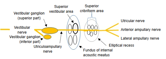
Inferior part of the vestibular ganglion
It gives two branches: one passes through the inferior vestibular area and the other through the foramen singulare to reach the vestibule.
1) Posterior ampullary nerve: through the foramen singulare to the sensory epithelium of the ampullary crest of the posterior semicircular duct.
2) Saccular nerve: through the inferior vestibular area to the sensory epithelium of the macula of the saccule.
- Cochlear nerve : small branches pass through the foraminous spiral tract to the spiral ganglion, whose cells supply the sensory epithelium of the spiral organ.
- Vestibular nerve
- Vestibular ganglion (superior part) : utriculoampullary nerve → utricular nerve, anterior ampullary nerve, lateral ampullary nerve
- Vestibular ganglion (inferior part) : posterior ampullary nerve, saccular nerve
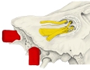 Nerves at the fundus
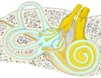 Vestibulocochlear nerve in the inner ear
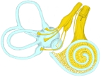 Cochlear and vestibular nerves
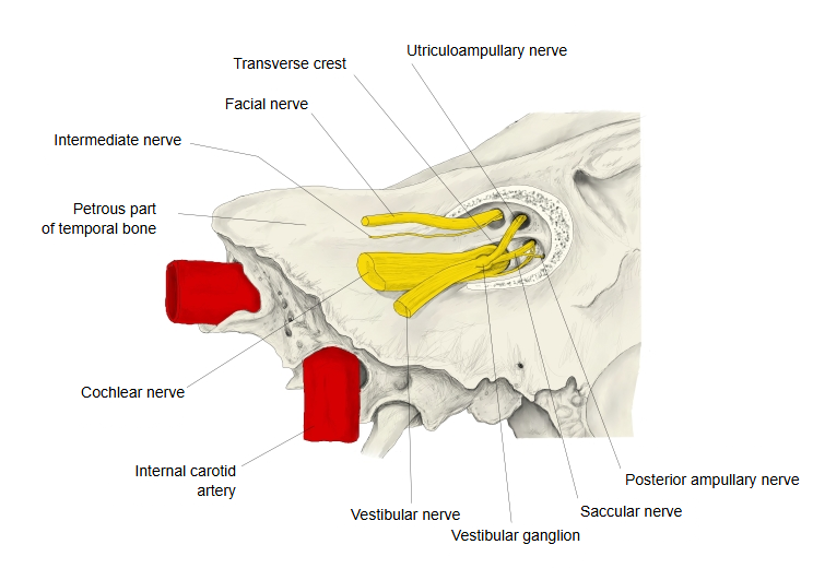
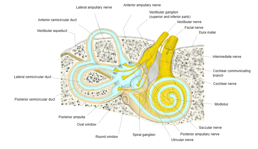
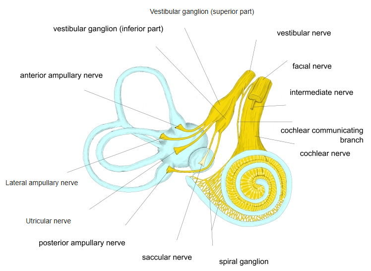
Reference websites
[Wikipedia]
· Vestibular ganglion, Wikipedia.
[Websites with illustrations]
· Nerves in the right internal acoustic meatus, with the vestibular (Scarpa's) ganglion (Medscape)
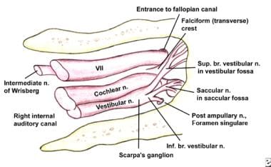
· Diagram of the four areas of the fundus of the internal acoustic meatus (Folia Morphologica)
Related terms
- Vestibular aqueduct
- External aperture of vestibular aqueduct
- Internal aperture of vestibular aqueduct
- Pyramid of vestibule
- Vestibular crest
- Scala vestibuli
- Vestibular nerve
- Vestibulocochlear nerve
- Fundus of internal acoustic meatus
- Internal acoustic meatus
- Vestibular maculae
- Ampullary crest
- Inferior cribriform area
References and sources
- Further reading: Vestibular nerve, Wikipedia.
- Federative International Programme for Anatomical Terminology (FIPAT). Terminologia Anatomica, 2nd ed. (TA2), 2019.